Dual Offer + FREE Dr Advice | WhatsApp Prescription | Book Home Visit ✆ 09220145540

Menu Icon

Osteosarcoma Diagnosis Management And Treatment

Osteosarcoma Diagnosis Management And Treatment

Posted By HealthcareOnTime Team Posted on 2022-03-24

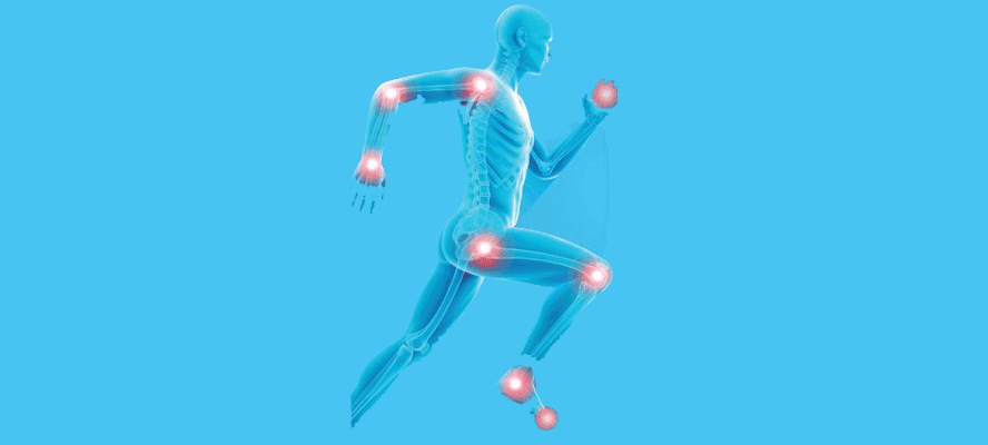
The very foundation of human body is defined by the bones. Strongest organ of all, providing support to all other organs in the body, along with producing white and red blood cells and storage of minerals, bones play a very significant and pivotal role in structuring the human body and aiding its motion. These sturdy bones can become vulnerable to many conditions associated with bone tissues and cells such as osteopenia, osteoporosis, osteomyelitis, osteomalacia, osteosarcoma, etc. A major disease associated with uncontrolled growth of bone cells is known as osteosarcoma on osteogenic sarcoma.

Osteosarcoma Diagnosis Management And Treatment

Osteosarcoma is one of the primary malignant bone tumour especially common in children and young adults (between 10 25 years), during period of adolescent growth spurt. This tumor of the skeleton is defined by formation of new immature bones or bone tissues by the tumor cells directly. Long bones are the primary regions of occurrence but osteogenic cancer can occur in any bone in the body. It is a condition characterized as highly cellular tumors of pleomorphic spindle shaped cells which can osteoid matrix (water, minerals, inorganic salts, collagen fibers, etc.). It rarely occurs in the soft tissues.

Characteristics of Osteosarcoma Prior to swelling of the soft tissues or enlargement of the bone mass, the patient might complain of pain which is an outcome of stretching of the periosteum. Pain can also arise as a result of bone weakening or development of minute fractures. The presence of tumor in the joint spaces also declines the range of motion, affecting their mobility.

Osteosarcoma develops due to rapid local growth of bone tissue which increases the likelihood of mutations that can ultimately develop into tumors. Aside from ionizing radiation as a probable cause, genetic predisposition to this Cancer is also likely. However, no DNA or RNA has been isolated for its cancer studies. Also, it has not been linked to chemical carcinogens or viruses.

Molecular Pathogenesis of Osteosarcoma
1. Bone Growth and Tumorigenesis Osteosarcoma occurs primarily in rapidly growing bones such as the distal femur, proximal tibia, or proximal humerus. Osteosarcoma mostly develops during puberty, when the epiphyseal growth plates are responsible for increase in height. Thus, the patients with this disease are taller as compared to normal individuals.

2. Chromosomal Abnormalities
DNA helicases separate the double strands of the DNA before replication, and patients with genetic mutations in RecQ helicase family are prone to higher risk of malignant tumors such as osteosarcoma. Other genetic mutations which are associated with osteosarcoma are, amplification of chromosome 6p21, 8q24, 12p14; loss of heterozygosity of 10q21.1, etc.
3. Tumor Suppressor Gene Dysfunction Tumor suppressor genes such as p53 and retinoblastoma (Rb) have a protective function against development of cancer due to environmental factors. Mutations in p53 and Rb genes result in loss of their function giving rise to malignant cell lines stimulating the pathogenesis of osteosarcoma.
4. Transcription Factors Functions such as cell proliferation, differentiation and bone metabolism are regulated by activator protein 1 complex (AP-1). This complex molecule is composed of two proteins Fos and Jun, whose expressions are elevated in case of high-grade osteosarcoma as compared to benign or low-grade tumors. Myc gene is a transcription factor responsible for cell growth and division. Overexpression of this gene is involved in resistance to chemotherapy and in pathogenesis of osteosarcoma.

Subtypes of Osteosarcoma
Osteosarcoma can be divided into three subtypes based on the microscopic features of the cells. The subtypes are as follows:
High-grade osteosarcomas
Of all the incidences of osteosarcoma in children and young adults, most are high-grade osteosarcomas. These cells when observed under microscope are unlike the normal bone cells and most of them are in the process of dividing into new cells. Some of all the fastest growing high-grade osteosarcomas are - osteoblastic, chondroblastic, fibroblastic, etc.
Intermediate-grade osteosarcoma and Low-grade osteosarcoma
Intermediate-grade falls below in between the high-grade and low-grade sarcoma and can mostly be considered as low-grade osteosarcoma. Periosteal (chondroblastic tumor composed of cartilaginous tissues arising from bone surface) is an example of low-grade osteosarcomas. Unlike high-grade, low-grade osteosarcoma are the slowest growing and the cells look normal with very few of them in the process of dividing. Parosteal (well differentiated fibroblastic tumor arising from surface of the bone) is one such type of osteosarcoma that falls in this category.
Tumor Staging
The staging of osteosarcoma is based on the size and location of the tumor, whether it has metastasized and to what extent. Staging of cancer helps to determine the severity, deduce survival statistics and plan treatment accordingly. Osteosarcoma can be classified into two broad groups: Localized and metastatic cancer.
Localized This kind of tumor has not spread and is observed to be present only at the site of origin i.e the bone it started in, and might extend to adjacent tissues of the bone. Easy to be excised by surgery.

Metastatic This kind of tumor spreads to other parts of the body such as the lungs (mostly), other bones, brain, etc. At this point, it becomes difficult to be removed by surgery.

Two types of system are referred for staging osteosarcoma
A. Musculoskeletal Tumor Society (MSTS) Staging System/Enneking System G1 - Low Grade
G2 - High Grade
T1 - Intercompartmental (within bone)
T2 - Extracompartmental (extended beyond the bone)
MO - Tumors not spread to lymph nodes or other organs
M1 - Tumor spread to lymph nodes and other organs

B. TNM Staging System
This staging system uses four important aspects in terms of T, N, M and G.
T Describes the size of the tumor along with its appearance in other areas of the bone.
N Describes the spread of tumor in proximity to the lymph nodes
M Describes the spread of the tumor to other distant organs of the body
G Describes the grade of the tumor specifying the microscopic features of the osteogenic cells.

Clinical Manifestations The first symptom is pain which wakes up the patient from sleep. The other signs and symptoms are as follows:
- Vascularity of tumors causing swelling and redness
- In case of metastasis to lung - cough, difficulty in breathing (dyspnea), chest pain may occur
- Limping or suffering from stress fractures (fracture caused by disease weakening the bones)

Diagnosis
A. Imaging Tests
1. Positron Emission Tomography (PET) Scan
A form of radioactive sugar (F"-FDG) is injected into the blood of the cancer patient. Because cancer cells grow rapidly, a large amount of tracer gets localized due to increased glycolysis. PET-CT imaging then reveals the sites in the body showing radioactivity due to increased uptake of radioactive glucose. PET scanning in diagnosis of osteosarcoma assist to stage the malignancies, to differentiate between fibrosis and recurrent tumor. In the presence of increased levels of tumor markers, PET scan can detect and localise tumor recurrence, to differentiate between benign and malignant tumors.

2. Radionuclide Bone Scan
Radionuclide bone scan is mostly recommended for detecting osseous (turned into bone or bone tissue) metastases. 20 mCi Tc" methylenediphosphate (TC." MDP) is injected intravenously and whole body images are captured after 3-4 hours. The distribution of the radiopharmaceutical is observed and areas with increased osteoblastic activity are noted and interpreted.

3. Magnetic Resonance Imaging (MRI) Scan
MRI is the modality of choice to see the local extent of the tumor due to its better spatial resolution. It has the ability to capture detailed images of the soft tissues in the body with the combination of radio waves and magnets, thus not involving radiations. For this technique, contrast material used is gadolinium- which is injected,intravenously. It's a very good modality to pick marrow metastases.

B. Biochemical Laboratory Tests
Biochemical blood tests are carried out after the diagnosis is done. Testing for elevated serum levels of biochemical markers such as alkaline phosphatase (ALP) and lactate dehydrogenase (LDH) assists in determining the level of advancement of osteogenic tumor.

C. Biopsy
Biopsy is a confirmatory test performed after complete examination and image testing. It is more specific for determining the type of tumor and its grade. Biopsy can be performed in two ways either by open or closed method.

1. Closed or Needle Biopsy
A small sample of tumor is extracted with the help of needle and biopsy is performed by the fine needle aspiration cytology (FNAC) and core needle technique.

2. Open or Incisional (Surgical) Biopsy
The tumor is extracted through the surgical cut on skin.

Management and Treatment
Treatment regimes differ for patients depending on the nature of the cancer found in the body i.e. whether the tumor is localized or metastasized. There are three categories based on which, management and treatment of osteosarcoma can be particularly specified, namely

Localized Osteosarcoma
For such cases, a combination of both chemotherapy and surgery to remove the tumor is followed, wherein chemotherapy may be carried out both before and after surgery. External radiation therapy may also be used for treatment. In this therapy, high-energy external radiation beams penetrates through the tissues, delivering the radiations into the areas where cancer cells are present. Complete surgical resection which may include amputation, or removal of tumor without amputation and replacement of bone and joints with prosthetic devices is important for complete cure.

Metastatic Osteosarcoma
Most common site of metastases is lung. If metastasectomy is possible, it is done. If not, then chemotherapy is considered.

Recurrent Osteosarcoma
Cancer reappearance in the treated patients is termed as recurrent osteosarcoma. Such patients should undergo surgery followed by post-operative chemotherapy. Most recurrences of osteosarcoma occurs in the lungs. Treatments are most effective when the diagnosis is carried out at earlier stages and the chances of complete recovery also rise with effective management.

The mere mention of the word cancer imbalances the state of mind but, to facing this hurdle determination to overcome is the key.


View Our Editorial Policy
Was this article helpful?

Trending Health & Fitness Web Stories

Find Latest Health Web Stories, Fitness Photo Stories, Health AMP Stories.VIEW ALL

Did you catch our latest post? JOIN US

Facebook LinkedIn Instagram Twitter YouTube

Contact Us

Email: info@healthcareontime.com | Phone No: 09220145540 | Whatsapp: 9820693367
  • Copyright 2024 HealthCareOnTime.com, All Rights Reserved
  • Disclaimer: HealthcareOnTime offers extensively researched information, including laboratory testing for health screening. However, we must emphasize that this content is not intended as a substitute for professional medical advice or diagnosis. Always prioritize consulting your healthcare provider for accurate medical guidance and personalized treatment. Remember, your health is of paramount importance, and only a qualified medical professional can make precise determinations regarding your well-being.
DMCA.com Protection Status HealthCareOnTime.com Protection Status HealthCareOnTime.com Protection Status