Dual Offer + FREE Dr Advice | WhatsApp Prescription | Book Home Visit ✆ 09220145540

Menu Icon

Immune Modulation In Respiratory Diseases

Immune Modulation In Respiratory Diseases

Posted By HealthcareOnTime Team Posted on 2022-02-02

Understanding Major Respiratory Diseases
Respiratory disease is one of the leading contributors to deaths in India and the world. India bears around 32% of the global burden of respiratory, diseases. In most cases, Smoking, lung infections, or genetic disorders playa vital role in major respiratory disease. India is one of the developing countries in which, both acute and chronic respiratory diseases are prevalent in substantial numbers. The major respiratory diseases which have maximum occurrence
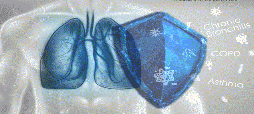
 in India are - Asthma,Chronic Obstructive Pulmonary Disease (COPD), Lung Cancer, Pneumonia, Pleural Effusion, Chronic Bronchitis, Tuberculosis, and Allergic Rhinitis. Based on the recent Global Burden of Disease (GBD) survey, among these diseases, asthma and COPD show off wide variations in several states in India with maximum affected people

Immune Modulation In Respiratory Diseases

Lung - A Critical Immune Interface
Lungs are the most crucial organs for vertebrates to survive, via the intake of oxygen from the air. Due to this continuous gaseous exchange with the air, lungs are most vulnerable to infections by toxins, microbes, airborne pathogens, or allergens. Based on the intensity and duration of exposure, lung infection can vary from mild to acute. For example, COPD and allergic asthma trigger chronic inflammatory responses in the lungs, whereas acute microbial infections cause pneumonia or sepsis with severe inflammation to the lungs which can lead to Acute Lung Injury / Inflammation(ALI) or Acute Respiratory Distress Syndrome (ARDS).

Being susceptible to such severe infections, lungs are designed to serve as a major immune organ that can protect themselves autonomously from the microbes, allergens, xenobiotics, or pathogens that enter our body through inhalation. Although, lung epithelial cells are the principal targets of most respiratory viruses, there is expanding evidence that lung epithelia themselves are capable of generating antimicrobial responses. This immuneagainst pathogens can also be enhanced by nasal adjuvant administration. Hence, lungs act as a potent immunological interface that can generate local as well as protective systemic immunity against external respiratory pathogens.

Major Immune Cells in Lungs
The figure below illustrates that lungs contain macrophages which may be divided into alveolar macrophages and interstitial macrophages. Lungs also contain alveolar and bronchial epithelial cells (AECs and BECs), Dendritic cells, Natural Killer cells along with other Innate Lymphoid Cells (ILCIS, ILC2s, and ILC3s), and adaptive immune cells like T and B cells to protect them against pathogens, toxicants, and allergens inhaled. Neutrophils also migrate to the lungs in response to infection or inflammatory insult. These pulmonary innate immune cells release several cytokines and, chemokines and serve as antigen presenting cells (APCs) to regulate pulmonary innate as well as adaptive immunity.

Know the Genes Causing Respiratory Diseases
There are multiple genes that play a vital role in the initiation and progression of lung diseases such as Tumor Necrosis Factor-a (TNFa), Transforming Growth Factor-B1 (TGFB1), Glutathione S-transferases P1 and M1 (GSTP1, and GSTM1), and superoxide dismutase 3 (SOD3). TLR3 and TLR5 trigger the progression of severe respiratory diseases like pneumonia and chemokine ligand 2, Toll-like receptor 4, T-helper cell 1 initiate tuberculosis. Researchers have further identified a gene that is closely associated with nicotine addiction and lung cancer. It has been seen that a specific region on chromosome 15 (15q25.1), containing the nicotinic acetylcholine receptor subunit genes CHRNA3 and CHRNA5 are responsible for the trigger point behind the lung carcinoma.

Pathogenesis of Respiratory Disease
Pathogenesis of major lung diseases depends on the type of infection and the area of exposure and are discussed below

1. Asthma As we know, asthma is a type of chronic inflammatory airway disease generally represented with the symptoms like - tightness in the chest, wheezing, coughing, and dyspnea. All these symptoms are generally associated with exacerbation of airway inflammation. Whatever be the stimulus the symptoms often start with smooth muscle contraction and inflammation.

Although for a long time, researchers have found asthma is a reversible health condition, recent research shows permanent structural changes in the airway such as - sub-basement membrane fibrosis, smooth muscle hyperplasia, new vessel formation, and glandular hyperplasia. All these changes are collectively known as airway remodeling which is a significant part of asthma pathogenesis. Research on persistent asthma pathogenesis showed that asthma airway remodeling causes an accelerated decline in lung function, which may be slower than COPD but its impact is permanent in bronchiolar airways.

2. COPD Bronchial biopsies and induced sputum of COPD patients have revealed the symptoms of lung inflammation. This inflammation appears to be an enhanced or abnormal inflammatory response, which is beyond the protective capacity of the lungs. It is suggested that the presence of increased CD8+ T lymphocytes differentiates between smokers who do and do not develop COPD. There's a correlation between T-cell numbers, the quantity of alveolar destruction, and therefore the severity of airflow limitation. However, smokers with normal lung function also show an increased number of CD8* cells compared to non-smokers. There's a significant decrease in T-lymphocyte infiltration in bronchial biopsy specimens from subjects with severe COPD. Other cells like macrophages, dendritic cells, epithelial cells, neutrophils play a vital role in the pathogenesis

3. Chronic bronchitis Inflammation of the central airways is a prominent feature in patients with bronchitis. The pathogenesis of bronchitis involves an inflammatory mononuclear cell infiltrate within the airway wall and a neutrophil influx into the airway lumen. The molecular events that trigger inflammation in mucus hypersecretion showed that chemotactic agents are derived not only from tissue fluid but also generated by the infected bronchial epithelium. For example - bronchial epithelial cells synthesize interleukin (IL-8), a potent chemoattractant and activator of neutrophils and lymphocytes. Airway inflammation often involves the adhesion of infiltrating leukocytes to resident parenchymal cells within the bronchi and to the extracellular matrix. The resultant inflammation likely plays an immediate role within the clinical features of the disorder.

Cells Involved in Pathogenesis
There are multiple cells that are involved in the pathogenesis of respiratory diseases

1. Mast cells The mast cells are one of the key players who trigger the early allergic response within minutes of antigen exposure. The mast cell surface-bound IgE is crosslinked by the antigen which leads to the activation of mast cells and potent mediators such as - histamine, leukotrienes, prostaglandin D2, thromboxane B2, and platelet-activating factors. These mediators result in the contraction of airway smooth muscle, edema, and enhanced mucous secretions which induce airflow limitation and generate acute asthma symptoms. Mast cells also contribute to the persistence of airway inflammation through cytokines (IFN-a, IL-1, IL-4, IL-5, IL-6, IL-8, IL-16) and chemokines (MIP-1a, MIP-1b, MCP, and RANTES) secretions and occurrence of latephase response.

2. Eosinophils The role of Eosinophils is closely connected to allergic diseases. They are often presented within the airways of allergic asthmatics and correlate with parameters of disease severity. These cells consist of potent mediators in their granules such as major basic protein, eosinophil cationic protein, eosinophil-derived neurotoxin, and eosinophil peroxidase. These proteins can induce airway damage and contribute to airway hyperresponsiveness; therefore, eosinophils have been proposed as principle effector cells in asthma pathogenesis. additionally, these cells, which include leukotrienes, cytokines, matrix metalloproteinase, and reactive oxygen species, have the capacity of generating other important factors that could contribute to airway obstruction and injury.

3. Neutrophils Recent studies that evaluated acute respiratory diseases (infectious also as noninfectious) have revealed the presence of increased number of neutrophils within the airway. These cells can presumably contribute to the pathogenesis of asthma, COPD, or bronchitis through the assembly of lipid mediators, reactive oxygen species, and proteases (myeloperoxidase and matrix metalloproteinase.

4. Lymphocytes Lymphocyte or T cells play an important role in the pathogenesis of asthma. A T cell subset, Th2 type, secrete cytokines like IL-4, IL-5, IL-9, and IL-13. These cells increase in number within the bronchoalveolar lavage fluid of atopic asthmatics following the introduction of an allergen within the airway. Through the generation of those cytokines, Th2 cells cause eosinophil activation, IgE production, mucus, and the expression of adhesion molecules like VCAM-1. The T cell is additionally capable of manufacturing numerousregulation of other inflammatory cells and therefore the worsening of acute and chronic inflammation.

5. Macrophages Macrophages are often found predominantly within the lower airway and represent almost 90% of cells recovered by bronchoalveolar lavage in normal and stable asthmatic patients. These resident cells significantly contribute to the normal host defense by phagocytosis, generation of enzymes, as well as reactive oxygen species. They also enhance the inflammatory response of cytokines like GranulocyteMacrophage colony stimulating factor (GM-CSF), IL-1, IL-6, lipid mediators (LTB4, C4, D4, PGD2, and thromboxane A2), and matrix metalloproteinase. Researchers have identified the significant ability of macrophages to get anti-inflammatory cytokines like IL10, IL-12, and TGF-b. A decrease in the production of those cytokines has been thought to trigger the upregulated airway inflammation in asthma. Also, alveolar macrophages can act as antigen-presenting cells within the airway.

6. Dendritic Cells Dendritic cells are considered as professional' antigenpresenting cells within the lung and have the potential to act against an antigen and present it to T lymphocytes with subsequent development of a Th2 cell type which will contribute to eosinophil maturation (through the generation of IL-5) and IgE production (through the generation of IL-4).

Immune Modulation with Current Therapy Over the past decade, there have been multiple trials of anti-inflammatory agents in patients who were suffering from severe pneumonia, developed sepsis, or systemic inflammatory response syndrome. The trials involved inhibitors of TNF and endotoxin. Though the trials fail to establish a potent therapy, it is now established that many of the cytokines, specifically TNF, are essential for host defense against both intracellular and extracellular pathogens. It gave a thought to the researchers that an infected organ would need to send signals to the bone marrow for a continued supply of neutrophils, for a sufficient host defense response.

On the basis of satisfactory preclinical data with G-CSF, clinical trials have been initiated with the patients of community-acquired pneumonia, multilobar pneumonia, and pneumonia with sepsis. A study suggested that G-CSF can be regarded as an effective immunomodulator in a variety of non-neutropenic patients such as hospital-acquired pneumonia or immunosuppressed by alcohol.

A recent study identified that faster radiological resolution in patients treated with G-CSF, accompanied with complications including adult respiratory distress syndrome and disseminated intravascular coagulation, G-CSF was well tolerated in a short trial of 18 patients with pneumonia and severe sepsis. These favorable trends trigger further studies in multilobar pneumonia and severe pneumonia with sepsis were initiated. Docke and colleagues investigated IFN-y_in patients with immunoparalysis associated with sepsis and revealed that downregulation of monocyte human leukocyte antigen (HLA)-DR expression identifies a subgroup of septic patients with a higher risk of hospital-acquired infection. Furthermore, the investigators showed that IFN enhanced HLA-DR expression, increased lipopolysaccharide-induced TNF responses, and improved clinical parameters of sepsis in more than 80% of patients treated with IFN.

Immunostimulants in Respiratory Diseases
Recently, there have been several treatments to showcase the incidence of ARTIS (Vitamin A, vitamin C, zinc, antibiotics). Among them, the trials with immunostimulants (herbal extracts, bacterial extracts, synthetic compounds) are very crucial which aim to enhance the immune defenses of the respiratory tract. However, the data quality of most of the studies was below the standard, insufficient, and too diverse to conclude.

Since chronic respiratory conditions remain global health threats, as exemplified in the Severe Acute Respiratory Syndrome Coronavirus 2 (SARS-CoV-2) pandemic. This manuscript explains that most of these chronic respiratory conditions are marked by underlying chronic inflammatory processes. Therefore, immune modulation has become a cornerstone for novel therapeutic options, shaping the future of pulmonary medicine. It is important to explore immune modulation as a promising option for future therapeutic interventions in respiratory diseases.


View Our Editorial Policy
Was this article helpful?

Trending Health & Fitness Web Stories

Find Latest Health Web Stories, Fitness Photo Stories, Health AMP Stories.VIEW ALL

Did you catch our latest post? JOIN US

Facebook LinkedIn Instagram Twitter YouTube

Contact Us

Email: info@healthcareontime.com | Phone No: 09220145540 | Whatsapp: 9820693367
  • Copyright 2024 HealthCareOnTime.com, All Rights Reserved
  • Disclaimer: HealthcareOnTime offers extensively researched information, including laboratory testing for health screening. However, we must emphasize that this content is not intended as a substitute for professional medical advice or diagnosis. Always prioritize consulting your healthcare provider for accurate medical guidance and personalized treatment. Remember, your health is of paramount importance, and only a qualified medical professional can make precise determinations regarding your well-being.
DMCA.com Protection Status HealthCareOnTime.com Protection Status HealthCareOnTime.com Protection Status