
cancer diagnosis is among the leading causes for morbidity and mortality in our country. Out of 1.2 billion Indians, around 1 million are diagnosed with cancer each year. The most common cancer in females is found to be breast cancer, followed by cervical cancer, while in males, the more common cancers are tobacco related. The rate of cancer incidence in India is lower as compared to developed nations, however, mortality rates in both developed nations and in India is almost comparable. This is because in many of these cases, mortality in cancer cases is due to low rates of early detection, less access to screening and poor treatment outcomes. Based on this, it is clear that there is a need for greater awareness regarding the technologies available to us in our fight against this disease.
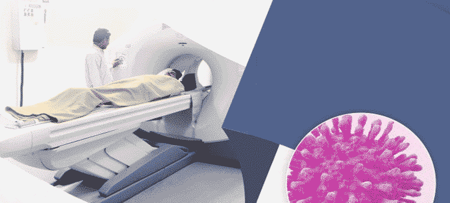
Cancer imaging is a collective term for the tests that can be used to scan the body for detection of tumors, how far these have spread and to observe the efficacy of the prescribed cancer treatment. In most of these tests, some form of energy (for instance, sound waves in an ultrasound, X-rays, radioactive particles or magnetic field in MRIS) is passed through the body. These forms of energy create an image of the internal tissues of the body which is studied by the doctor to look for cancer.
Imaging has many applications in cancer treatment and monitoring, it can be used to detect cancer when it has not produced symptoms, as being in the early stages. These techniques can also be used as a part of cancer screening tests. In other cases, it is done to locate the tumor if a person is presenting with symptoms of cancer. Cancer imaging can also help in the staging of cancer, to plan treatment, and to determine if the tumor has responded to the treatment. It can also provide evidence of cancer recurrence.
PET-CT-A Promising Tool
Conventionally, MRI and CT scans have been used for many years to identify and
classify tumors based on their anatomy, shape and size, density, and water content. More recently,
Positron emission tomography-computed, tomography (PET-CT) has been developed which is a cancer
imaging modality that combines the PET and CT techniques. While PET provides information regarding
metabolic changes in particular tissues, CT imaging allows precise detection of the anatomical region
where the abnormal Metabolic changes have occurred. Thus, a PET-CT scan creates pictures of the body
in which abnormally acting tissues can be observed. It can characterize tumors based on biochemical
changes at the molecular level. The first PET-CT scanner in India was inaugurated at the Tata Memorial
Center in December 2004.
Positron Emission Tomography (PET)- The Workings PET
uses molecules labelled with radioisotopes like “c, “N, “O, “F and “Ga which are injected into
the body and release positrons. These positrons travel a certain distance dissipating the kinetic
energy in excitation/ionization interactions and come to rest. Positron undergoes mutual
annihilation with a nearby electron. The resting masses are converted into a pair of 511 keV photons
that are emitted simultaneously in 180?? opposite directions. This can be detected by the PET scanner
which then identifies the location within tenths of a millimeter to few mm where these radiolabelled
isotopes have accumulated.
One of the characteristics of cancer cells that distinguish it from normal body cells is that they have greater tendency to take up and breakdown glucose to meet their substantial energy needs- this is called as Warburg effect. Currently, PET uses fluorine-18fluorodeoxyglucose (FDG) as the radiolabel due to its similarity to glucose, which has a high rate of uptake in a wide majority of tumors. Based on the isotope uptake by different regions of the body, an image of the body can be generated. Some of the cancers most commonly imaged with FDG PET include lymphoma, squamous cell carcinoma of head and neck, colorectal cancer, breast cancer, and melanoma. PET can also be used for differentiation of benign from malignant tissue.
FDG-The wonder molecules
The wide adoption of FDG as the radiolabel can be attributed to the fact that FDG is excreted in the urine
and is hence rapidly cleared from the body. Further, its halflife of 110 minutes allows it to be transported
from the site of production to the testing facility with ease.
PET-CT scanning involves intravenous injection of FFDG in concentrations based on the patient’s body weight. The patient should be in fasting for 6 to 7 hours prior to the procedure. This is followed by intravenous administration. After 1 hour, the patient is positioned on the scanner and the test is run.
Computed Tomography (CT-The Augmenting Technology
The size of the tumor, and its precise location in the body cannot be accurately measured by a PET
scan alone. This can be done by CT scan which also helps in staging the cancers and detecting its
spread to nearby tissues. The change in tumor size is still used to monitor tumor response to therapy.
A CT scan uses X-rays to take a series of pictures from different angles and combines them to create
cross-sectional images of bones, tissues and blood vessels. These tissues can be distinguished from
each other based on their density. An advancement of the CT technique is the use of spiral CL,
wherein the X-ray beam moves in a spiral path with no gap in between the images. CT can also identify
specific features or phenotypes of tumors (such as its texture), which can be further used to learn about
the vascularity of the tumor. Thus, it also informs about the prognosis, response to anti-angiogenic drugs
and makes predictions about drugi delivery to tumors.
PET-CT: In the Current Scenario
The increase in the use of PET-CT over other imaging technologies is driven by several factors, foremost
among which is its higher sensitivity and specificity for the detection and staging of tumors, as compared
to the conventional imaging methods. So far, PET-CT has been used for staging of several cancers with high
accuracy, such as non-small cell lung cancer, lymphoma, breast cancer, esophageal cancer, melanoma,
and head and neck cancers. It can also be used to characterize the tumor. PET-CT has been reported to
accurately detect distant metastases and secondary cancers.
The anatomical features of the tumor which can be studied by PET-CT help in pre-surgical decision making regarding surgical interventions, radiation therapy, or biopsy planning. Since PET-CT delivers greater accuracy in tumor staging, it can help identify which patients would benefit from surgery against those who would not. Also, in radiation therapy, PET-CT can help provide better target definition and guide radiologists to the biopsy site. Based on the uptake of FDG, PET-CT images can give insight into tumor proliferation activity, aggressiveness and prognosis,
Treatment Response Assessment
F-FDG has been successfully used to assess tumor response to tumorocidal and targeted therapy,
such as cytostatic therapies- the response is promptly visible as a decrease in the uptake of FDG by
the tumor cells in response to the therapeutic agent. Tumor response to treatment as early as after
a single cycle of chemotherapy has been assessed by this technique. PET-CT images have the potential
to guide biopsies of the most metabolically active regions of the tumor, and it can be used to modulate
the dose of radiation therapy, although monitoring radiation treatment is slightly more difficult with
PET-CT since FDG uptake is unchanged immediately after treatment. “F-FDG PET-CT has also been
investigated as a prognostic marker for prediction of outcomes. “F-FDG PET-CT can determine the
efficacy of the treatment by detecting changes in tumor biology within days or even hours of starting
therapy. This makes it powerful for tumor response and also allows for chemotherapies to
be tailored based on the response of that particular patient.
Future Prospects. The Journey Ahead
Aside from imaging the tumour itself, PET-CT has also been reported to be effective in monitoring abnormal
cellular proliferation of the cancer cells, imaging of hypoxia (which can lead to cellular changes in the tumor
resulting in more aggressive phenotypes), and for the imaging of apoptosis. The latter has the potential to
give an early indication of the success of therapy. Using PETCT to detect abnormal cellular proliferation has
already shown promising results in its ability to predict tumor grade in lung cancers, evaluate brain tumors
and can serve as predictor of tumor response. Additionally, PETCT can monitor protein synthesis and cell
membrane metabolism. Since protein production is up-regulated in cancer cells, PET-CT using radiolabelled
amino acids
can be used for tumor imaging. Finally, tracers that target intracellular and cell-surface receptors
which are unique to or are overexpressed in cancer cells have also been developed for use in PET-CT.
Precision Medicine with PET-CT
Precision Medicine with PET-CT” Precision medicine involves the use of therapies that are specifically
tailored to act on particular molecular targets expressed by the disease or tumor. This can further allow
greater personalization of therapeutic approaches for individual patients. Progress in the discovery of
targeted therapeutic agents has opened the gates of oncology to precision medicine, which has had
significant role in minimizing over and under treatment, thereby preventing unnecessary toxicity,
morbidity and therapy failure. There are several requisites in the application of precision medicine to
oncology, in each of which PETCT can play an important role.
Patient Selection based on biomarkers
It is important to identify the right biomarkers that can
distinguish between patients who will benefit from a particular therapy, against those who would not.
In such situations, PET-CT comes into play through the use of radiopharmaceuticals that have specific
molecular targets and which can determine the efficacy of the targeted therapy. PET-CT can therefore,
be coupled with therapeutic agents to provide a combined diagnostictherapy approach for precision medicine.
PET-CT against tumor heterogeneity
All tumor cells in an individual’s body are not alike at the molecular level; different masses may
have different propensity for metastasis even when they appear histologically similar. Even within
the same tumor, different cells may have different features which present a challenge in development
of targeted therapy, if tumor biology is gained by sequencing cells from one portion of the tumor
(sampling error). Also, in some cases, the tumor may be located at regions that are hard to reach for
biopsy. Tumor heterogeneity can be explored using PET-CT both for cells within the same tumor,
or between primary tumor and metastases, all in a single study. This is possible due to the availability
multiple PET radiopharmaceuticals.
Early therapy assessment to identify resistance to therapy
Identifying therapy resistance early is important in precision medicine, and PET-CT helps
with that as well. PET-CT can be used to identify the molecular subtype of the tumor, since,
in some cancers such as head and neck squamous cell cancers, the affinity of the tumor cells
for FDG before and after therapy changes drastically and can be monitored to determine if the
therapy is working. Interim FDG PETCT after 2 cycles of chemotherapy in Hodgkin’s
lymphoma is another clinical example .
It is important to time the PET-CT procedure correctly, with respect to therapy cycles. Standardized methods for PET CT calibration, patient preparation, image acquisition and image analysis are also essential for precision therapy assessment. Finally, there is need for developing novel therapy assessment methods using FDG PET-CT or other pharmaceuticals in the near future, since radiation therapy induced inflammation can interfere with the intensity of FDG uptake; this can lead to errors in therapy assessment.
In the future, PET reporter gene imaging is expected to be used for trafficking of cell-based therapies. Additionally, pharmacokinetic studies done by PET can also help in improving drug development. Characteristics perfusion and texture will be surmised from dynamic CT images, which can be used to indicate tumor vascularization As newer tracers for PET are being made available for routine clinical use, the potential of PET-CT is on the rise for more promising clinical applications. The ultimate goal is to improve diagnostic accuracy, treatment, Surveillance and prognosis of cancer.