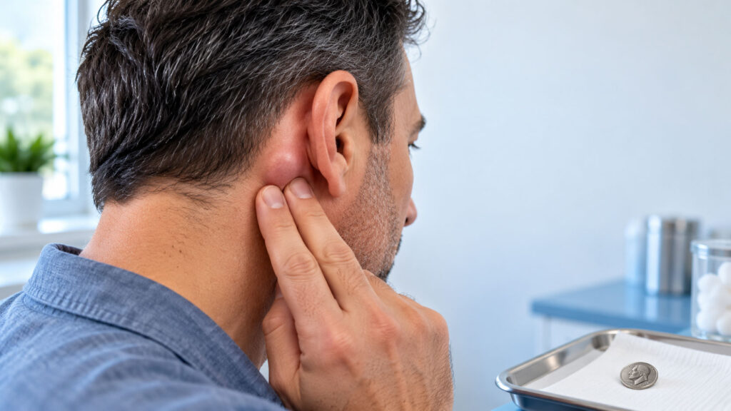
Here’s something most people get backward. You find a lump behind your ear, notice it doesn’t hurt, and feel relieved.
Table of Contents
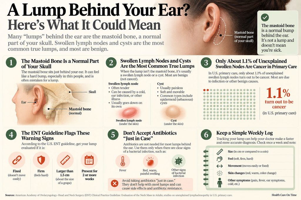
With lumps, pain is often the reassuring sign. Sore, tender lumps usually mean your body is fighting a simple infection. The lump doctors watch more closely is the quiet one: hard, fixed, and not going away.
That doesn’t mean yours is serious. Most aren’t. It means “no pain” isn’t a reason to ignore it, and a simple 2-week rule tells you when to act.
Quick Answer: A hard, painless lump behind the ear is usually harmless: a swollen lymph node, a skin cyst, or normal bone. See a doctor if it lasts 2 weeks or more without shrinking, grows past about 1.5 cm (a bit over half an inch), feels firm or stuck in place, has broken skin over it, or comes with fever, night sweats, weight loss, hearing changes, or trouble swallowing.
At a Glance
- Many “lumps” behind the ear are the mastoid bone, a normal part of your skull.
- Swollen lymph nodes and cysts are the most common true lumps, and most are benign.
- In US primary care, only about 1.1% of unexplained swollen nodes turn out to be cancer.
- The US ENT guideline flags lumps that are fixed, firm, larger than 1.5 cm, or present 2+ weeks.
- Don’t accept antibiotics “just in case” unless there are clear signs of infection.
- A simple weekly log (size, feel, movement, skin, symptoms) can speed up your diagnosis.
What That Hard Lump Behind Your Ear Probably Is
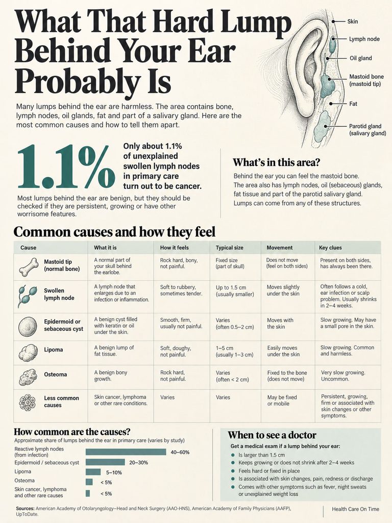
The area behind your ear is crowded. It holds bone, lymph nodes, oil glands, fat, and the edge of a salivary gland.
That’s why one small bump can have several explanations. Here are the usual suspects, starting with the most common and least worrying.
First, Rule Out Normal Anatomy
Press firmly behind your earlobe, a little down and back. You’ll likely feel a hard, rounded knob of bone. That’s the mastoid tip, part of your skull’s temporal bone.
Everyone has one on each side. Patients who reach out to us often discover it for the first time while scratching an itch or adjusting their glasses, and assume it’s new. Quick check: if you feel the same bump on the other side, it’s almost certainly normal bone.
Swollen Lymph Node (Posterior Auricular Node)
You have small lymph nodes just behind each ear, called posterior auricular nodes. They filter fluid from your scalp and the skin around your ear.
When they react to a scalp rash, dandruff flare, insect bite, ear infection, or cold, they can swell to pea size or a bit larger. The American Academy of Family Physicians treats nodes larger than about 1 cm as enlarged.
A reactive node often feels rubbery, moves a little under your finger, and shrinks over 2 to 4 weeks. It can stay small and firm for months after an infection, which is usually fine.
Epidermoid or Sebaceous Cyst
This is a small sac under the skin filled with keratin, a soft protein from skin cells. It feels smooth, round, and firm, and it moves with the skin when you push it.
Some have a tiny dark dot (a pore) on top. They grow slowly, stay painless, and only turn red and sore if they get infected or rupture.
Lipoma
A lipoma is a benign lump of fat. It’s usually soft and doughy, not hard, and slides easily under the skin.
If your lump is truly hard, a lipoma is less likely. Still, a lipoma sitting deep or near tight tissue can feel firmer than expected.
Osteoma and Other Bony Growths
An osteoma is a benign bone growth on the skull. It’s rock hard, doesn’t move at all, and grows very slowly over years.
It feels fixed because it’s part of the bone. That feeling can be alarming, but osteomas are not cancer and often need no treatment unless they bother you.
Less Common Causes Worth Knowing
A small number of hard, painless lumps need a closer look:
- Skin cancer. Ears and the skin behind them get a lot of sun. Basal cell or squamous cell skin cancer can show up as a firm bump, scaly patch, or sore that won’t heal.
- Parotid gland lump. Your largest salivary gland wraps around the front and bottom of the ear. A lump low near the earlobe or jaw angle may come from it. Most parotid tumors are benign, but all need evaluation.
- Lymphoma. A cancer of immune cells that can cause firm, rubbery, painless nodes, sometimes in several places.
- Metastatic node. Cancer from elsewhere in the head, neck, or scalp spreading to a nearby node. This one tends to be hard, fixed, and growing.
Our medical reviewers see the same pattern again and again: the lump people fear most is rarely the one they have. Here’s how the main causes compare.
| Cause | How it feels | Moves? | Typical size | Usual timeline | Next step |
| Mastoid tip (normal bone) | Rock hard, smooth | No | Same on both sides | Always been there | No action needed |
| Reactive lymph node | Rubbery, firm | Slightly | 0.5 to 1.5 cm (pea to dime) | Shrinks in 2 to 4 weeks | Track for 2 weeks |
| Epidermoid cyst | Smooth, firm, round | Moves with skin | 0.5 to 3 cm | Slow growth over months | Routine visit if it grows or bothers you |
| Lipoma | Soft, doughy | Yes, easily | 1 to 5 cm | Very slow growth | Routine visit |
| Osteoma | Rock hard | No, fixed to bone | 0.5 to 2 cm | Slow growth over years | Routine visit, imaging if unsure |
| Skin cancer | Firm, scaly, or sore | Moves with skin | Any size | Doesn’t heal in 4+ weeks | Dermatologist |
| Lymphoma or metastatic node | Hard or rubbery, may be fixed | Often no | Over 1.5 cm | Grows or stays 2+ weeks | Prompt doctor visit |
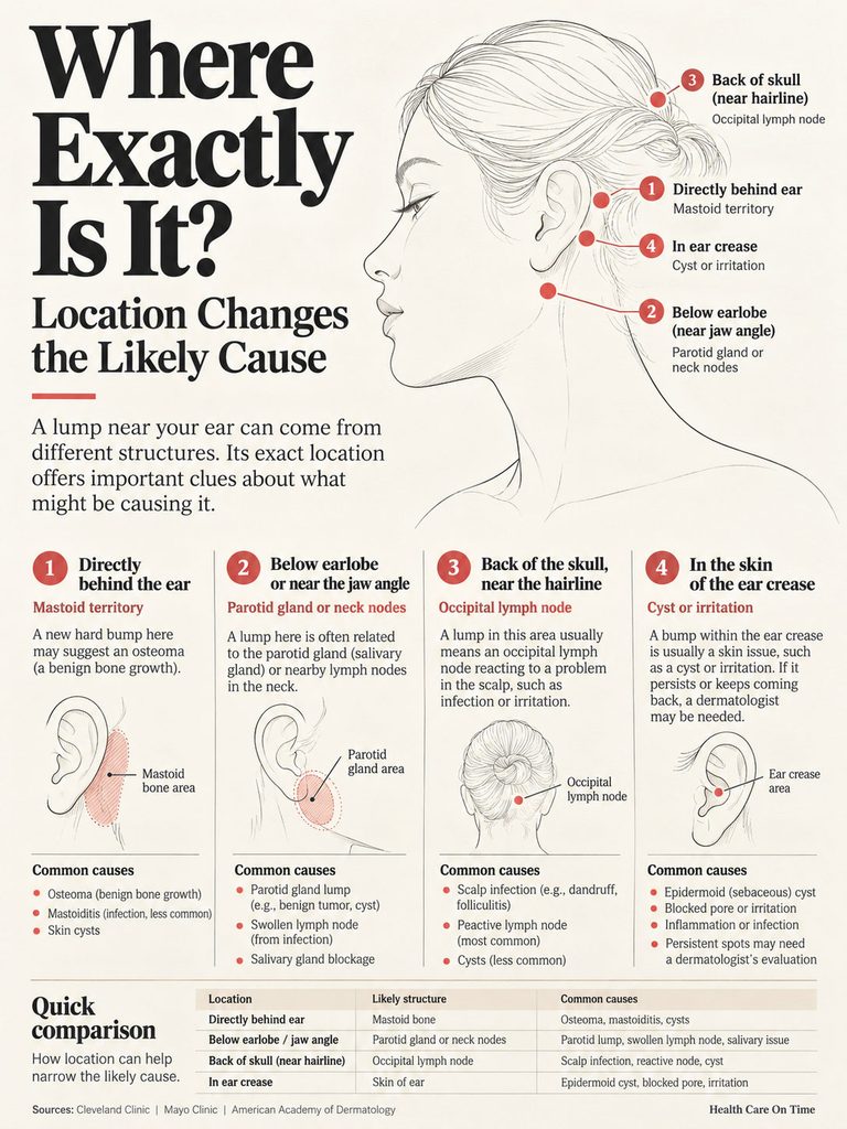
Where Exactly Is It? Location Changes the Likely Cause
“Behind the ear” covers more ground than most people realize. Pinpointing the spot helps you and your doctor narrow things down fast.
Directly Behind the Ear, on the Bone
This is mastoid territory. A hard, smooth bump here that matches the other side is normal bone. A single, rock-hard, fixed bump that’s new may be an osteoma. Posterior auricular nodes also sit here, just under the skin.
Below the Earlobe or Near the Jaw Angle
This spot sits over the lower part of the parotid gland and the upper neck nodes. Lumps here are more likely to be salivary gland growths or neck nodes linked to the throat, tonsils, or teeth. This is the area where the ENT guideline matters most.
Back of the Skull, Near the Hairline
A bump lower and farther back, near where the skull meets the neck, is often an occipital lymph node. These react to scalp problems like dandruff, lice, eczema, or a recent haircut nick.
In the Skin of the Ear Crease
A bump right in the fold where the ear meets the head is usually a cyst, a blocked pore, or irritation from glasses arms or mask straps. A scaly spot that won’t heal here needs a dermatologist.
Why “Painless” Is Not the Same as “Harmless”
Pain usually comes from inflammation. Infected cysts, abscesses, and nodes fighting a sore throat swell fast and hurt.
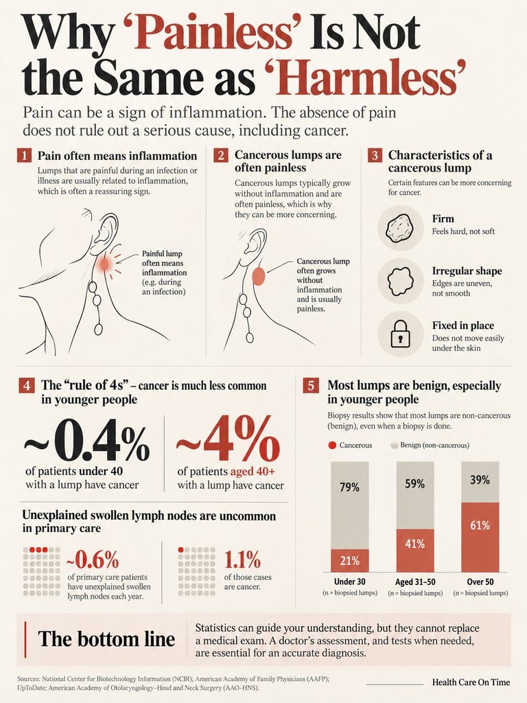
That’s why a painful lump that appeared during a cold tends to be reassuring. It has an obvious cause, and it usually fades as you recover.
What a Worrying Lump Tends to Feel Like
Lumps from cancer usually grow without much inflammation, so they often don’t hurt. According to Medical News Today, a cancerous lump is typically firm, irregular in shape, and fixed in place.
“Fixed” means it doesn’t slide when you push it gently side to side. It feels anchored to deeper tissue. The exception is bone: an osteoma or the mastoid tip also feels fixed, but it’s smooth and has usually been there a long time.
The Real Odds, by Age
Here’s the good news in hard numbers. In primary care, unexplained swollen lymph nodes show up in about 0.6% of patients each year, and only 1.1% of those cases are cancer, according to AAFP.
Age changes the picture. Cancer was found in about 4% of patients 40 and older with unexplained nodes, versus 0.4% of those under 40. Doctors call this the “rule of 4s.”
Biopsy data point the same way. In one series of 628 patients who had a node sampled, benign causes were found in 79% of those under 30, 59% of those aged 31 to 50, and 39% of those over 50 (AAFP). These were people already sent for biopsy, so real-world odds for a new lump are far lower.
What the Numbers Can’t Tell You
Be honest with yourself about three limits. First, the 1.1% figure comes from older Dutch primary care data, still the best available but not US-specific or recent.
Second, those numbers cover swollen nodes in general, not lumps behind the ear alone. Third, no statistic replaces an exam. Low odds are a reason to stay calm, not a reason to skip the visit when the 2-week rule says go.
The 2-Week Rule: When to See a Doctor
The American Academy of Otolaryngology (the main US ENT society) published a clinical guideline on neck lumps in adults. It’s written for doctors, but its rules translate well for you.
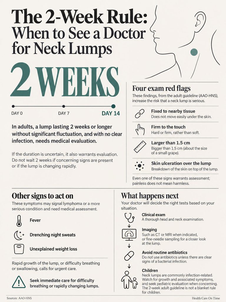
The core idea: an adult neck lump that isn’t clearly from infection and sticks around needs a proper workup. The area behind and below your ear counts.
The Time Rule
The guideline treats a lump as higher risk when it has been present 2 weeks or more without real change, or when you can’t say how long it’s been there (AAO-HNS).
The ENT society’s patient material adds two related warning signs: the lump keeps getting bigger, or it shrinks but never fully goes away. A node from an infection should disappear completely once the infection clears.
The Four Exam Red Flags
According to the same guideline, any one of these features raises the risk:
- Fixed to the tissue underneath (doesn’t slide)
- Firm or hard consistency
- Larger than 1.5 cm, about 0.6 inch, a little smaller than a dime
- Ulcerated skin over it (an open sore or broken skin)
A hard, painless lump already ticks one box. That’s why the 2-week clock matters more for you than for someone with a soft, tender bump.
Symptom Red Flags
Book a prompt visit if the lump comes with any of these, all listed in the ENT guideline’s patient summary:
- Fever over 101°F
- Unexplained weight loss
- Drenching night sweats
- Voice changes or hoarseness
- Trouble or pain with swallowing
- Hearing loss or ear pain on the same side
- Ongoing throat or neck pain
Fever, night sweats, and losing more than 10% of your body weight without trying are classic warning signs for lymphoma, per AAFP.
Go Today: Urgent Signs
Get same-day care or go to the ER if you have:
- A red, hot, swollen area behind the ear that pushes the ear forward (possible mastoiditis)
- High fever with severe ear pain or drainage
- New weakness or drooping on one side of your face
- A fast-growing lump that makes breathing or swallowing hard
Children Are Different
The adult guideline doesn’t apply to kids. In children, infection causes most neck lumps, and small nodes are very common.
Call your pediatrician if a child’s lump keeps growing, gets larger than a nickel, lasts more than a few weeks without shrinking, or comes with fever, weight loss, or night sweats.
Why You Shouldn’t Accept Antibiotics “Just in Case”
The ENT guideline advises against routine antibiotics for a neck lump unless there are signs of bacterial infection, such as redness, warmth, tenderness, or fever.
Why it matters: an antibiotic trial can cost weeks. If the lump shrinks a little, everyone relaxes, and a real diagnosis gets delayed. Patients who book visits through us are often surprised to learn they can politely ask, “Are there signs of infection here, or should we image it first?”
What to Track: A Simple Weekly Lump Log
Most advice says to “monitor” the lump without explaining how. Doctors find a good log more useful than any description given from memory.
How to Measure Without a Ruler
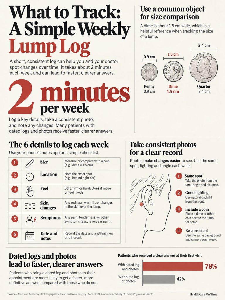
Compare the lump to common objects. Use the same one each week.
- Pencil eraser: about 0.25 inch (6 mm)
- Pea: about 0.4 inch (1 cm)
- Dime: 0.7 inch (about 1.8 cm)
- Nickel: 0.84 inch (about 2.1 cm)
- Quarter: 0.96 inch (about 2.4 cm)
The key line is 1.5 cm. If your lump is close to the size of a dime or bigger, it has crossed the guideline’s size threshold. A soft tape measure works too: gently feel for the edges and measure across.
The 6 Things to Log Each Week
- Date you first noticed it, and today’s date
- Size compared to a pea, dime, nickel, or quarter
- Feel: soft, rubbery, firm, or rock hard
- Movement: slides easily, moves a little, or doesn’t move
- Skin: normal, red, scaly, dimpled, or open sore
- Other symptoms: fever, night sweats, weight change, ear or throat problems, recent cold, scalp rash, new medicine
Write it in your phone’s notes app. It takes about 2 minutes.
How to Take Comparable Photos
Ask someone to take the photo, since you can’t see behind your own ear. Use the same spot, lighting, and angle each week.
Place a coin next to the lump for scale. Daylight from the front works best. Light from the side can make a small lump easier to see.
What Not to Do
- Don’t squeeze or try to pop it. Cysts can rupture and get infected under the skin.
- Don’t use needles or pins at home.
- Don’t press on it many times a day. Repeated poking can irritate it and make it feel bigger.
- Don’t apply wart removers or acids. They can damage skin and hide a skin cancer.
- Don’t sit on a “maybe” for months. If you reach the 2-week mark with no shrinking, book the visit.
Across patients we serve, those who arrive with a dated log and two or three photos tend to get clear answers faster, often with fewer repeat visits.
Who Is at Higher Risk
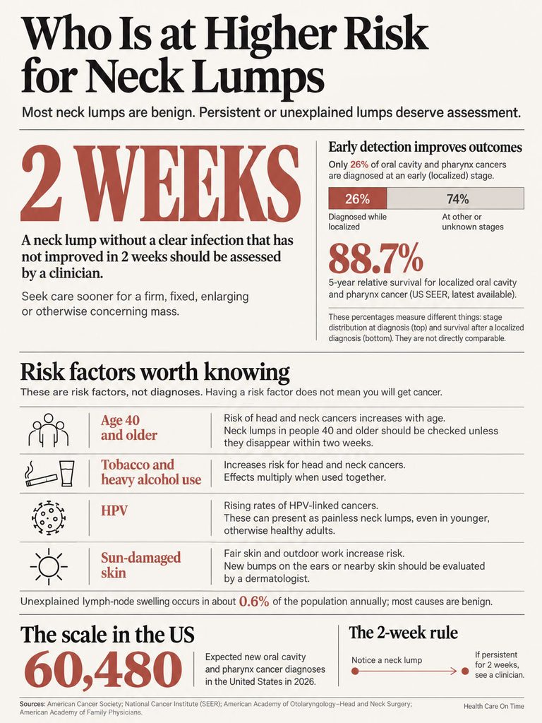
Most people with a lump behind the ear have nothing serious. These factors make a careful workup more important, not more frightening.
Age 40 and Older
AAFP lists age over 40 as a key risk factor, along with male sex and symptoms like fever, night sweats, and weight loss. A head and neck surgeon quoted by AARP says a lump near the ear in an older adult needs checking unless it disappears within a week or two.
Tobacco and Heavy Alcohol Use
Smoking, vaping, chewing tobacco, and heavy drinking all raise the risk of head and neck cancers. Together, they multiply it.
HPV and Younger Adults
The old picture of a neck cancer patient was an older smoker. That’s changing. The American Cancer Society reports that oral cavity and oropharyngeal cancer rates have risen steadily since the mid-2000s, largely due to HPV-linked cancers.
These cancers often show up first as a painless neck lump in otherwise healthy adults, many of whom never smoked. The HPV vaccine helps prevent them.
Sun-Damaged Skin
The tops and backs of the ears get heavy sun and are often missed by sunscreen. If you have fair skin, work outdoors, or have had skin cancer before, any new bump here deserves a dermatologist’s look.
Other Factors
- A weakened immune system (HIV, transplant medicines, chemotherapy)
- A recent cat scratch or bite (cat-scratch disease can swell nearby nodes)
- Certain medicines that can enlarge nodes, including allopurinol, carbamazepine, and phenytoin (AAFP)
Here are the US numbers that put the risk in perspective.
| Statistic | Figure | Source |
| Unexplained swollen lymph nodes in primary care (per year) | About 0.6% of patients | AAFP, American Family Physician 2016 |
| Unexplained nodes that turn out to be cancer | 1.1% overall | AAFP, American Family Physician 2016 |
| Cancer risk with unexplained nodes, by age | 0.4% under 40 vs 4% at 40 and older | AAFP, American Family Physician 2016 |
| Adults with palpable neck nodes on routine exam | 56% in one primary care study | AAFP, American Family Physician 2002 |
| New oral cavity and pharynx cancers expected in the US | 60,480 cases | American Cancer Society, 2026 estimates |
| New Hodgkin lymphoma cases expected in the US | About 8,920 (average age at diagnosis: 39) | American Cancer Society, 2026 |
| Trend in new oral cavity and pharynx cancer rates | Up about 1.0% a year (2013 to 2022) | NCI SEER |
| 5-year survival when oral and pharynx cancer is found early (localized) | 88.4% | NCI SEER |
That last number is why the 2-week rule exists. Early detection changes outcomes. Yet according to SEER, only about 26% of these cancers are caught while still localized.
What Happens at the Doctor’s Office
Knowing what to expect makes the visit less stressful and more useful.
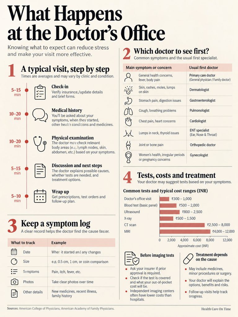
Which Doctor to See First
- Primary care doctor: The best first stop for most adults. They can examine you, order tests, and refer you.
- Dermatologist: A good first choice if the lump is clearly in the skin, scaly, or sore-like.
- ENT (otolaryngologist): The specialist for lumps that are deep, fixed, near the parotid gland, or flagged as higher risk. Many PPO plans let you book directly; HMOs usually need a referral.
The Exam and Questions
Your doctor will feel the lump and check your ear canal, eardrum, throat, mouth, scalp, and other node areas (neck, collarbone, armpits). Expect questions about:
- When you noticed it and whether it has changed
- Recent infections, dental work, or scalp problems
- Tobacco, alcohol, and sun exposure
- Fever, sweats, weight loss, or fatigue
- Medicines and pets
This is where your log pays off. Show the dates, sizes, and photos.
Tests You Might Need
- Neck ultrasound: Shows whether a lump is solid or fluid-filled and checks its shape. Often the first look for cysts and nodes in lower-risk adults.
- CT or MRI with contrast: Detailed images of deep tissue and bone. The ENT guideline recommends this for adults at higher risk.
- Fine needle aspiration (FNA): A thin needle collects cells for testing. The guideline prefers it over cutting the lump out as a first biopsy.
- Blood tests: A complete blood count (CBC) and sometimes inflammation markers, especially with fever, fatigue, or several swollen nodes.
One more point from the guideline: a lump that looks like a cyst on imaging still needs follow-up until there’s a clear diagnosis in higher-risk adults. Some cancers spread to nodes that look cystic.
Rough US Costs and Insurance Notes
Prices vary widely by state, facility, and plan. These are rough self-pay ranges, not quotes:
- Office visit: $100 to $300
- Neck ultrasound: $200 to $600
- CT neck with contrast: $500 to $3,000 (hospitals often charge more than imaging centers)
- FNA biopsy with lab testing: $400 to $1,500
Ask your insurer whether imaging needs prior approval. If you’re paying cash, ask for a self-pay price before booking. Independent imaging centers are often far cheaper than hospitals.
Treatment by Cause
- Normal mastoid bone: No treatment.
- Reactive lymph node: Watch and wait; treat the infection behind it, if any.
- Cyst: Leave it if small; remove it fully if it grows, gets infected, or bothers you. Draining alone often lets it come back.
- Lipoma or osteoma: Removal only if it grows or causes problems.
- Skin cancer: Usually removed in the office or with Mohs surgery.
- Lymphoma or other cancer: Referral to a specialist team. Outcomes are best when it’s found early.
Your Next Step: A Quick Decision Guide
Find the row that matches your lump right now. In cases reviewed by our medical team, this simple sorting answers most people’s “go now or wait?” question.
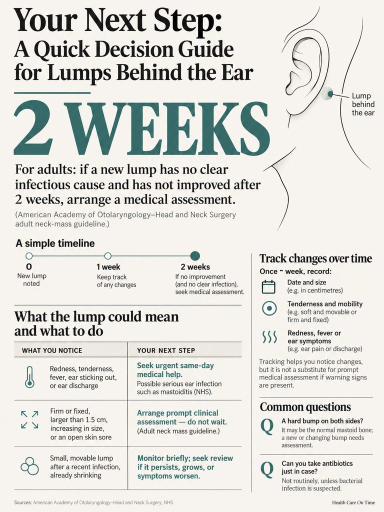
| Scenario | What it may mean | Recommended action |
| Hard bump on both sides, behind and below the ears, always there | Normal mastoid bone | No action needed |
| Pea-sized, rubbery, appeared during a cold or scalp rash | Reactive lymph node | Log weekly; see a doctor if not shrinking at 2 to 4 weeks |
| Smooth, round, moves with skin, may have a dark pore | Epidermoid cyst | Routine visit; don’t squeeze it |
| Lump present 2+ weeks with no shrinking, or start date unknown | Needs evaluation per ENT guideline | Book a primary care visit within 1 to 2 weeks |
| Firm, fixed, or dime-sized (1.5 cm) or larger | Higher-risk features | Prompt visit; ask about imaging |
| Lump plus fever, night sweats, or unexplained weight loss | Possible lymphoma or body-wide illness | See a doctor within days |
| Scaly bump or sore that won’t heal in 4 weeks | Possible skin cancer | Dermatologist visit |
| Red, hot swelling pushing the ear forward, with fever | Possible mastoiditis | Same-day care or ER |
Frequently Asked Questions
Should you worry about a hard lump behind your ear that doesn’t hurt?
Not right away, but don’t ignore it. Most are cysts, reactive nodes, or normal bone. Because hard, painless lumps can occasionally be serious, track it for 2 weeks. If it hasn’t shrunk, is 1.5 cm or larger, or feels fixed, see a doctor for an exam and possibly imaging.
Is it normal to feel a hard bone behind your ear?
Yes. The mastoid tip is a hard, rounded part of the skull behind and slightly below each earlobe. Everyone has one. If you feel a matching bump on the other side and it has always been there, it’s almost certainly normal anatomy and needs no testing.
How long can a swollen lymph node behind the ear last?
Most reactive nodes shrink within 2 to 4 weeks after an infection clears. Some stay small and firm for months, which is often fine. A node that keeps growing, passes 1.5 cm, or doesn’t shrink after 2 to 4 weeks without a clear cause should be checked.
What does a cancerous lump behind the ear feel like?
It’s usually hard or firm, irregular in shape, and fixed in place, meaning it doesn’t slide when pushed. It often doesn’t hurt and tends to grow over weeks. Feel alone can’t confirm cancer, though. Only imaging and a biopsy can give a definite answer.
How can you tell a cyst from a lymph node at home?
A cyst usually sits in the skin, moves with the skin, and may show a tiny dark pore on top. A lymph node sits slightly deeper, feels rubbery, and often appears after a cold or scalp irritation. Home checks are only a guide; an exam or ultrasound confirms it.
Can a lump behind the ear be lymphoma?
It can, but it’s uncommon. Lymphoma nodes tend to be firm, rubbery, painless, and sometimes appear in several places, like the neck or armpits. Fever, drenching night sweats, and unexplained weight loss raise suspicion. A biopsy is the only way to diagnose it.
What kind of doctor should check a lump behind your ear?
Start with a primary care doctor, who can examine it and order tests. See a dermatologist if it’s clearly a skin growth or sore. An ENT specialist handles deep, fixed, or higher-risk lumps and lumps near the salivary gland below the ear.
Can stress or a cold cause a lump behind the ear?
A cold, sore throat, ear infection, or scalp rash can easily make nearby lymph nodes swell. Stress itself doesn’t create lumps, but it can trigger scalp or skin flare-ups that do. A node from a cold should fully disappear within a few weeks of recovery.
Is a pea-sized lump behind the ear serious?
Usually not. Pea-sized (about 1 cm) nodes and cysts are common and mostly harmless. Track it weekly. See a doctor if it grows toward dime size, becomes harder or fixed, lasts beyond 2 to 4 weeks without shrinking, or comes with fever, sweats, or weight loss.
Should you try to pop or drain a lump behind your ear?
No. Squeezing a cyst can rupture it under the skin and cause a painful infection. Poking a lymph node or bony lump does nothing useful and can irritate the area. If a cyst bothers you, a doctor can remove it fully so it’s less likely to return.
Do you need an ultrasound for a lump behind your ear?
Not always. If the lump is clearly a cyst or small reactive node, an exam may be enough. Ultrasound helps when the cause is unclear. For higher-risk adults, the US ENT guideline recommends contrast CT or MRI, often followed by a needle biopsy.
Is a lump behind the ear in a child more or less worrying?
Generally less. Kids get swollen nodes often because they catch many infections. Still, call a pediatrician if the lump keeps growing, gets larger than a nickel, lasts more than a few weeks, or comes with fever, weight loss, night sweats, or red swelling behind the ear.
Disclaimer: This article is for general education and has been reviewed by the HealthCareOnTime medical team. It is not a diagnosis or a substitute for professional medical care. If you have a new or changing lump, see a licensed healthcare provider. For urgent symptoms such as high fever, facial weakness, or trouble breathing or swallowing, call 911 or go to the nearest emergency room.
References
- Pynnonen MA et al. Clinical Practice Guideline: Evaluation of the Neck Mass in Adults. Otolaryngology–Head and Neck Surgery, 2017 (PubMed)
- AAO-HNSF: Neck Mass Guideline Press Release and Fact Sheet
- Gaddey HL, Riegel AM. Unexplained Lymphadenopathy: Evaluation and Differential Diagnosis. American Family Physician, 2016
- Bazemore AW, Smucker DR. Lymphadenopathy and Malignancy. American Family Physician, 2002
- American Cancer Society: Estimated New Cancer Cases and Deaths, 2026
- American Cancer Society: Key Statistics for Oral Cavity and Oropharyngeal Cancers
- American Cancer Society: Key Statistics for Hodgkin Lymphoma
- National Cancer Institute SEER: Oral Cavity and Pharynx Cancer Stat Facts
- Medical News Today: What Causes a Lump Behind the Ear?
- AARP: 5 Possible Causes for Lumps Behind the Ears