You glance down and there’s a scatter of tiny red dots on your forearm. They weren’t there yesterday. Your brain reaches for the easy answer: razor burn, bug bites, maybe a reaction to a new detergent.
Table of Contents
That instinct to wave it off is exactly the one doctors warn against. Clinicians have seen petechiae written off as ordinary rash, only for a blood panel to reveal a problem hiding underneath.
Most of the time, those small red spots really are nothing. Occasionally they’re the body raising a flag. The good news is you can usually tell which is which in about ten seconds, using a test you can run right now at your kitchen counter.
Quick Answer: Tiny red dots on the skin are usually either petechiae or cherry angiomas. Petechiae are flat, pinpoint spots from bleeding under the skin that do not fade when pressed, and they can signal low platelets or infection. Cherry angiomas are harmless, slightly raised red growths that often blanch (lighten) under pressure and need no treatment. The press test, plus a quick check for fever or rapid spreading, tells you whether to relax or call a doctor.
At a Glance
- Petechiae are flat and non-blanching; cherry angiomas are raised and often blanch.
- The press test (glass test) is the fastest way to separate them at home.
- Cherry angiomas are extremely common with age and are benign.
- Petechiae that appear suddenly, spread fast, or come with fever need urgent care.
- About 50% of US adults over 30 have at least one cherry angioma.
- When in doubt, a quick doctor visit or a simple blood test settles it.
What Are Those Tiny Red Dots on Your Skin?
Red dots on the skin mostly fall into two camps that look alike but behave nothing alike. Telling them apart starts with knowing what each one actually is.
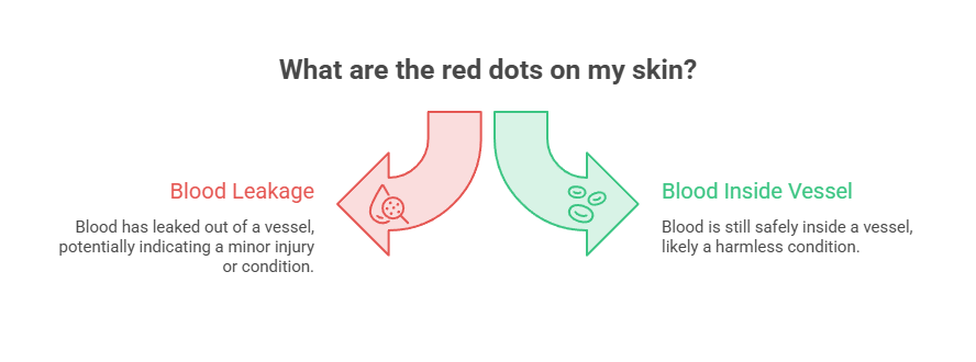
One comes from blood that has leaked out of a vessel. The other comes from blood that’s still safely inside one. That single distinction drives almost everything that follows, including whether you should worry at all.
Petechiae: Bleeding Under the Skin
Petechiae are tiny, flat spots, usually 2 millimeters or smaller, that form when small blood vessels called capillaries break and leak a bit of blood beneath the skin’s surface. The Cleveland Clinic describes them as pinpoint dots caused by this minor bleeding.
The color runs from red to purple to brownish. They often show up in clusters, sometimes dozens at once over a small patch, and they tend to appear suddenly rather than creeping in over years.
Because the blood has already escaped the vessel and pooled in the surrounding tissue, pressing on petechiae won’t push it away. That’s why they hold their color under pressure, a clue we’ll lean on heavily in a moment.
Petechiae can also shift color like a fading bruise as they heal, moving from red toward brown or yellow. They turn up anywhere, though the arms, legs, chest, and stomach are the usual neighborhoods. They can even appear on the inside of the mouth or the lining of the eyelids.
Cherry Angiomas: Harmless Blood Vessel Growths
Cherry angiomas are small, bright red growths built from a cluster of dilated blood vessels sitting just below the skin. They usually measure 1 to 5 millimeters and often look slightly raised, like a smooth, rounded dome.
Doctors also call them Campbell de Morgan spots or senile angiomas. They cluster most often on the chest, stomach, back, and arms, and they multiply as you age. They rarely turn up on the face, hands, or feet.
Here’s the reassuring part. Cherry angiomas are benign. They aren’t cancer, and across the cases reviewed by our medical team, a typical cherry angioma almost never points to anything dangerous on its own.
Early cherry angiomas can start out flat and barely noticeable, then slowly rise into the classic domed shape. Once they form, they’re permanent and won’t fade away, which is one quiet way to separate them from spots that come and go.
Why These Two Get Confused
Both are small. Both are red. Both can appear without warning. At a glance, a single small cherry angioma and a lone petechia can look nearly identical, which is why people swap the two constantly.
The real differences hide in the fine print. Whether the spot is flat or raised, whether it blanches under pressure, and whether it travels solo or in a crowd. Nail those three and you’ve mostly solved it.
What Red Dots Look Like on Different Skin Tones
Red spots don’t read the same way on every complexion. On deeper skin tones, both petechiae and cherry angiomas can look darker, leaning purple, brown, or nearly black rather than bright red, which makes them easier to overlook.
Dermatology references note that cherry angiomas tend to stand out more on lighter skin. On richer skin tones, the flat-versus-raised check and the press test carry even more weight, since color alone becomes a less reliable guide. Good lighting and a careful fingertip pass help a lot here.
Petechiae vs Cherry Angioma: The Differences That Matter
Placed side by side, the two conditions separate fast. This is the table worth screenshotting before your next mirror check.
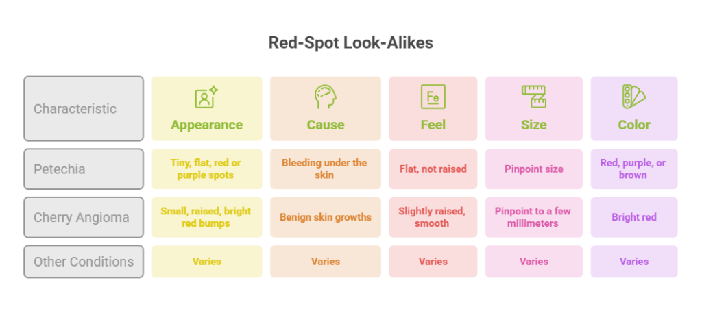
| Feature | Petechiae | Cherry Angioma | Why It Matters |
| Size | 2 mm or smaller | 1 to 5 mm | Petechiae tend to be smaller and more uniform |
| Texture | Flat, can’t feel them | Slightly raised, smooth dome | Run a fingertip lightly across the spot |
| Color | Red, purple, or brown | Bright cherry red | Petechiae darken and fade as they heal |
| Pattern | Clusters, appear suddenly | Scattered, build over years | Many spots at once leans toward petechiae |
| Press (blanch) test | Do not blanch | Often blanch or lighten | The single most useful at-home clue |
| Seriousness | Can signal an illness | Almost always harmless | Decides whether to watch or call a doctor |
The blanch row is the heart of the whole comparison. A spot that stays stubbornly red under pressure behaves differently from one that lightens, and that behavior reveals where the blood is sitting.
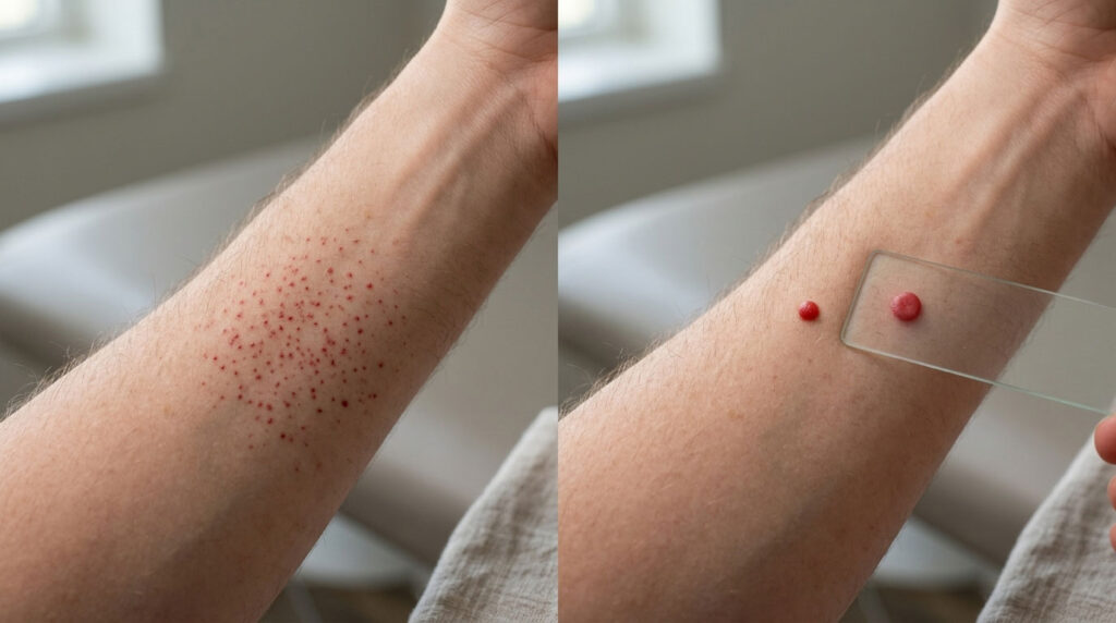
The 10-Second Press Test You Can Do at Home
The quickest way to sort red dots is the blanch test, also called the press test or glass test. It checks whether the redness comes from trapped blood (petechiae) or from blood still flowing inside a vessel (cherry angioma).
Patients booking tests through HealthCareOnTime often ask us how to run this correctly without overthinking it, so here’s the clean version.
How to Do the Glass Test Step by Step
- Find good light and locate the red spots clearly.
- Press a clear drinking glass (or a fingertip) firmly over them for a few seconds.
- Watch the skin directly under the pressure.
- If the spots fade or lighten, they blanch, which points toward a cherry angioma or a common rash.
- If the spots stay the same color while you press, they’re non-blanching, which points toward petechiae.
One firm caution. The press test is a useful starting clue, not a diagnosis. As one skin comparison guide notes, that test alone isn’t enough to rule everything out.
Non-blanching spots paired with fever, fast spreading, or simply feeling unwell always deserve prompt medical attention. The next sections lay out exactly when to make that call.
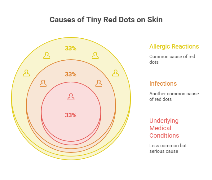
What Actually Causes Tiny Red Dots on the Skin?
Petechiae and cherry angiomas come from very different places. Cherry angiomas are mostly about aging vessels and genetics. Petechiae are about something making capillaries leak in the first place.
Sorting the cause matters because it changes your next move. One group of causes calls for a shrug; the other calls for a phone call. Knowing which bucket you’re in is half the battle.
Common, Harmless Causes
Plenty of everyday events rupture tiny capillaries. Hard straining during vomiting, violent coughing, heavy lifting, or childbirth can all do it, according to the Cleveland Clinic. You might notice these petechiae on the face or neck after a rough coughing fit.
Minor trauma works the same way. A tight tourniquet during a blood draw, a baby’s hard crying spell, even aggressive scrubbing in the shower. These petechiae usually stay local and fade on their own within several days.
For cherry angiomas, the drivers are mostly age, genetics, and hormones. Pregnancy can bring a fresh batch on, and a family history of the spots raises your odds of developing them.
Sun exposure may contribute in some cases too. None of these everyday causes signals disease, which is why a stable, slowly growing collection of red spots rarely worries clinicians. A spot that’s been quietly sitting on your back for two years is the picture of harmless.
Serious Causes That Need a Doctor
Sometimes petechiae are the first visible sign of a deeper issue. Our medical reviewers stress that the spots themselves aren’t the threat; the underlying cause is what matters.
Conditions that can produce petechiae include low platelet counts (thrombocytopenia), certain infections, blood cancers, and autoimmune disease like lupus. Liver disease and serious vitamin deficiencies can play a part as well.
The numbers explain the caution. According to Healthline, 50% to 60% of people with meningitis develop petechiae, and the spots are also a classic early warning sign of leukemia. Meningitis, in particular, can move quickly, which is why a fever-plus-spots combination is treated so seriously.
The specific conditions behind worrying petechiae are worth knowing by name. Meningitis and sepsis are the urgent infections; leukemia and immune thrombocytopenic purpura (ITP) sit among the blood disorders that drop platelet counts.
Others include lupus, chronic liver disease, severe vitamin C or vitamin K deficiency, and mononucleosis, the viral infection common in teens and young adults. Henoch-Schonlein purpura, an inflammation of small blood vessels, mainly affects children and often arrives with belly pain and joint aches.
Petechiae, Low Platelets, and What They Signal
Platelets are the cells that help your blood clot. When they drop too low, capillaries leak more easily and petechiae scatter across the skin. This is the mechanism behind most serious petechiae.
In leukemia, abnormal cells crowd out the bone marrow that makes platelets. As Moffitt Cancer Center explains, the body then can’t reliably plug broken capillaries, so blood escapes toward the surface and forms tiny red dots.
There’s an encouraging note here. Specialists at Roswell Park point out that petechiae fade once the underlying cause is found and treated. Other platelet-related triggers include immune thrombocytopenic purpura (ITP), certain medications (some antibiotics, antidepressants, and blood thinners), and vitamin C deficiency, which leads to scurvy.
It Might Be Neither: Red-Spot Look-Alikes
Not every small red spot is a petechia or a cherry angioma. Several other conditions show up looking similar, and confusing them is easy even for careful, attentive people.
Knowing the impostors helps you dodge both false alarms and false comfort. Here are the ones that surface most often in real life.
Spider Angiomas
A spider angioma has a central red dot with thin vessels radiating outward like spider legs. Press the center and the legs briefly vanish, then refill once you let go.
A cluster of these on the face or upper chest, especially in someone who drinks heavily, can point toward liver disease, as noted in this dermatology guide. A single one on a child, by contrast, is usually no cause for alarm.
Blood Blisters
A blood blister is a raised pocket of blood, usually from a pinch, rub, or repeated friction. The giveaway is timing. You can often recall the exact moment it happened, and it tends to fade over a week or two.
Cherry angiomas don’t fade on their own and have usually been there a while. If you can trace a spot back to last Tuesday’s pinch, it’s almost certainly a blood blister, not an angioma.
Pyogenic Granulomas
A pyogenic granuloma is a fast-growing red bump that can resemble a large cherry angioma. The difference is speed and behavior. These grow rapidly, bleed easily, and often develop after a minor skin injury.
Because they bleed so readily and grow so quickly, they usually warrant a doctor’s evaluation. A spot that balloons in size over days, rather than sitting unchanged for months, fits this pattern, not a typical cherry angioma.
Heat Rash, Folliculitis, and Keratosis Pilaris
Heat rash brings tiny red bumps to sweaty, friction-prone areas. Folliculitis centers red spots around hair follicles, sometimes capped with a small whitehead.
Keratosis pilaris creates rough, chicken-skin bumps on the backs of the arms and thighs. All three usually blanch and feel different from flat petechiae under a fingertip.
Across patients we serve, these everyday rashes get mistaken for petechiae far more often than any serious cause does. Texture is your ally: if you can feel raised bumps, you’re almost certainly not looking at true petechiae.
How Common Are These Red Spots, Really?
If you’ve just found a cherry angioma, you’re in enormous company. The data shows how routine these spots become once people move past their twenties.
| Statistic | Figure | Source |
| US adults over 30 with a cherry angioma | About 50% | Cleveland Clinic |
| Adults over 75 with cherry angiomas | About 75% | Cleveland Clinic / DermNet |
| Adolescents with at least one | About 5% | DermNet |
| Typical petechiae size | 2 mm or less | Healthgrades / PubMed |
| Purpura size (the larger relatives) | 4 to 10 mm | GoodRx |
| Meningitis patients who develop petechiae | 50% to 60% | Healthline |
The cherry angioma figures come straight from the Cleveland Clinic and DermNet, which both report the spots becoming near-universal in older adults. Even teenagers develop them on occasion.
The takeaway is simple. Cherry angiomas are an ordinary feature of aging skin, while petechiae are the spots that earn a closer look. When someone in their sixties finds a few new red dots that have been slowly accumulating, the odds heavily favor harmless angiomas rather than anything worrying.
It’s also worth knowing that petechiae have larger cousins. When the leaked-blood spots grow past about 4 millimeters, doctors call them purpura, as GoodRx explains. Larger non-blanching marks follow the same get-it-checked logic as petechiae.
When Cherry Angiomas Appear Suddenly
Most cherry angiomas creep in gradually over years, but some people notice a small burst of several at once. Hormonal shifts, including pregnancy, are a common trigger for this kind of sudden appearance.
A rapid crop of many new angiomas, sometimes called eruptive cherry angiomas, is usually still harmless. In rare cases, dermatology references link a sudden eruption to other internal changes, so a fresh wave of spots is worth a quick mention to your doctor rather than a quiet wait.
When to Worry: Red Flags You Should Never Ignore
Here’s where the press test hands off to judgment. Certain combinations of symptoms move red dots from watch-and-wait to get-help-now, and recognizing them quickly can matter a great deal.
| If You Notice… | It Might Mean… | What to Do |
| Spots with fever, stiff neck, or confusion | Possible meningitis or sepsis | Call 911 or go to the ER now |
| Spots spreading fast over hours | Serious bleeding or infection | Seek emergency care immediately |
| Spots with easy bruising or bleeding gums | Low platelets | See a doctor within 1 to 2 days |
| Sudden crop of non-blanching dots, no clear cause | Needs blood work | Book a doctor visit promptly |
| Raised red spot, stable for months, blanches | Likely a cherry angioma | Routine; mention it at your next visit |
| A spot that bleeds repeatedly or changes shape | Needs evaluation | Schedule a dermatologist check |
Call 911 or Go to the ER Now
Some signs simply can’t wait. Healthline lists urgent warning signs including spots that spread or grow, trouble breathing, a racing heart, unusual sleepiness, and other bruising appearing alongside the dots.
A fever with non-blanching spots in a child is a medical emergency until a doctor says otherwise. Because meningitis can progress fast, this combination always means immediate care rather than a wait-and-see approach. Trust your gut here, especially with kids.
See a Doctor Within a Few Days
Less urgent, but still important. A fresh batch of petechiae with no obvious cause, petechiae alongside easy bruising or frequent nosebleeds, or spots that keep returning after they fade.
In cases reviewed across our diagnostic network, a simple complete blood count (CBC) frequently explains new petechiae within a day. That’s why our medical team steers people toward testing rather than guessing, since a blood panel turns uncertainty into a clear answer you can act on.
Keep a quick mental log too. Note when the spots appeared, whether they’re spreading, and any other changes like fatigue, frequent infections, or unusual bleeding. That short history helps your doctor zero in on the cause faster at the visit.
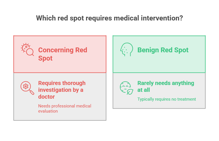
How Red Dots Are Diagnosed and Treated
What happens at the doctor’s office depends on which type of spot you have. One needs investigation; the other rarely needs anything at all.
How Doctors Diagnose Petechiae
A doctor usually opens with a visual exam, then asks about recent illness, current medications, and any straining or injury. This history alone often narrows the cause right away.
From there, blood tests check your platelet count and overall blood health. If results look off, more targeted testing follows, sometimes including a referral to a hematologist or other specialist.
The goal is always to find and treat the root cause, because the petechiae themselves clear up once that cause is handled. A doctor may also look for related clues like anemia, swollen lymph nodes, or signs of infection while putting the picture together.
Depending on the suspected cause, testing can extend to clotting studies, vitamin levels, or screening for infection. If a platelet problem looks likely, the workup sometimes includes a closer look at how the bone marrow is producing blood cells to pin down why platelets have dropped.
Cherry Angioma Removal Options and Typical US Costs
Cherry angiomas don’t need treatment from a medical standpoint. Removal is a cosmetic choice, or a practical one if a spot keeps snagging on clothing and bleeding.
Common in-office options in the US include electrocautery (burning the vessels with a fine probe), laser therapy, and shave removal. Costs vary widely by clinic and the number of spots, often running from roughly $100 to $400 or more per session, and insurance usually won’t cover purely cosmetic removal.
Patients commonly ask us whether they can handle this at home. The honest answer is no. DIY removal risks bleeding, scarring, and infection, while a dermatologist can take care of it safely in just a few minutes.
Recovery is quick. Most removal sites heal within a week or two, sometimes leaving a small flat mark that fades with time. A reputable dermatologist will walk through scarring risk and the small chance a spot grows back before treating it.
Can You Prevent Them?
You can’t reliably prevent cherry angiomas. Age and genetics drive them, and there’s no proven way to stop new ones from forming. The best approach is simply monitoring rather than prevention.
For petechiae, prevention means addressing the cause. Managing platelet conditions, reviewing medications with your doctor, and getting enough vitamin C all help. Protecting skin from hard friction and trauma reduces the minor, mechanical petechiae that follow straining or pressure.
Gentle skin habits help at the margins too. Easing up on harsh scrubbing, treating a cough that lingers, and staying on top of nutrition won’t guarantee spotless skin, but they trim the everyday capillary stress that brings on the harmless kind of red dots.
What to Do Right Now (Your Action Plan)
Found a tiny red dot and not sure what to think? Run this quick checklist before you spiral or shrug it off.
- Do the press test. Note whether the spot blanches or stays put under pressure.
- Check for company. Is it one stable spot, or a sudden cluster that appeared together?
- Scan for red flags. Fever, spreading, bruising, or simply feeling unwell changes everything.
- Watch harmless-looking spots for a few days. Cherry angiomas stay put, while minor petechiae fade.
- When anything feels off, or you just want certainty, see a doctor or book a blood test.
Frequently Asked Questions
Why do I suddenly have tiny red dots on my skin?
Sudden red dots are often petechiae from broken capillaries after coughing, vomiting, or straining, or they may be new cherry angiomas. A sudden cluster that doesn’t blanch, especially alongside fever or bruising, should be checked by a doctor promptly rather than ignored.
Are tiny red dots on the skin always serious?
No. Most tiny red dots are harmless, especially stable cherry angiomas or minor petechiae from straining. Concern rises when spots appear suddenly in clusters, don’t blanch when pressed, or arrive with fever, bruising, or feeling unwell. Those combinations warrant prompt medical attention.
Do cherry angiomas blanch when you press them?
Cherry angiomas often blanch or lighten partly when pressed, since they hold blood that’s still inside vessels. Very vascular ones may blanch only slightly. Petechiae, by contrast, don’t blanch at all, because the blood has already leaked into the surrounding skin tissue.
Are petechiae a sign of leukemia?
Petechiae can be a sign of leukemia, but they aren’t specific to it. Leukemia lowers platelets, so capillaries leak and tiny red dots form. Many other causes exist too. New, unexplained petechiae with fatigue, bruising, or frequent infections should be evaluated by a doctor quickly.
What vitamin deficiency causes red dots on the skin?
A vitamin C deficiency, known as scurvy, can cause petechiae along with swollen gums, joint aches, and easy bruising. It’s uncommon in the US but still occurs. Restoring vitamin C through diet or supplements typically resolves it once a doctor confirms the underlying cause.
How do I tell petechiae from a heat rash?
Heat rash usually brings raised, sometimes itchy bumps in sweaty areas, and it tends to blanch when pressed. Petechiae are flat, pinpoint, and don’t blanch. If you can feel the bumps and they fade under pressure, heat rash is far more likely than petechiae.
Do cherry angiomas mean I have liver problems?
Cherry angiomas don’t signal liver problems. You may be thinking of spider angiomas, which have a central dot with radiating vessels. Multiple spider angiomas on the chest or face, especially with heavy alcohol use, can point toward liver disease and deserve a medical check.
Can stress or straining cause red dots?
Physical straining can. Hard coughing, vomiting, heavy lifting, or childbirth can rupture tiny capillaries and produce petechiae, often on the face, neck, or chest. Emotional stress alone doesn’t directly cause them, though it can worsen conditions that do. These mechanical petechiae usually fade within days.
How long do petechiae take to go away?
Petechiae from minor straining or trauma often fade within a few days to two weeks as the trapped blood reabsorbs. Petechiae caused by an underlying condition clear only once that cause is treated. Spots that linger or keep returning should be evaluated by a doctor.
Should I remove a cherry angioma?
Removal is optional, since cherry angiomas are benign. Many people simply leave them alone. Consider removal if a spot is cosmetically bothersome or keeps catching and bleeding. A dermatologist can remove it safely with laser or electrocautery; avoid any do-it-yourself removal to prevent scarring and infection.
Are red dots on the skin a sign of diabetes?
Red dots aren’t a typical sign of diabetes itself. However, diabetes can affect small blood vessels and skin over time, and certain related skin changes occur. Petechiae specifically point more toward platelet or vessel issues. Persistent or unexplained spots are always worth discussing with your doctor.
When should I take my child to the ER for red spots?
Take your child to the ER right away if non-blanching red spots appear with fever, a stiff neck, drowsiness, trouble breathing, or rapid spreading. These can signal meningitis or a serious infection. When in doubt with a child’s non-blanching rash, seek emergency care without delay.
Medical Disclaimer: This article is for general informational purposes only and is not a substitute for professional medical advice, diagnosis, or treatment. Always consult a qualified healthcare provider about any skin changes or symptoms you notice. If you see non-blanching red spots accompanied by fever, spreading, or other warning signs, seek medical care promptly.