Genetic Screening
Screening basically means to assess an asymptomatic person for the risk of unsuspected disease or
disorder, to ensure the healthy status of that person. Genetic Screening tests are also done to evaluate
the risk of having a child with certain disorder. Screening usually involves Newborn screening (NBS),
preconception or reproductive genetic screening, and family history assessment.
Screening Vs Diagnosis
Screening tests are carried out to detect potential disease indicators in absence of symptoms, Whereas
Diagnostic Test are performed to establish the presence or absence of disease mostly following the
display of certain sign and symptom. Screening tests measure the degree or level of risk but cannot be
considered as a confirmatory test, while diagnostic test is the definite test which can determine the
defects in chromosome leading to various genetic disorders,
Sensitivity and specificity act as a measure to assess the validity of both screening and diagnostic tests. Sensitivity of a test is the ability to correctly classify a diseased individual as diseased while specificity is the correct identification of the individual as disease-free. They are calculated with the help of formulas and are usually inversely proportional i.e. higher the sensitivity lower is the specificity. Screening tests are more inclined towards being sensitive than specific. Hence, such tests declares majority of disease free patients with a possibility of having disease. Therefore, diagnostic tests are carried out for further investigation. It is considered as a good option to primarily screen the patients with a high sensitive low specific test and later with a test that is low sensitive but highly specific.
Not a fault in the stars, But a fault in the genes, An extra chromosome or a base pair shift Seems minute yet enough to label misfit, Can averting this faulty inheritance mean giving a new lease? Can timely screening prove us acquittal from the disease..
Genetic constitution of an individual can be said as the mirror which reflects a person’s characteristics. This reflection is controlled by gene expression which is the association between genetic information and the physical manifestation. Genetics controls the majority of aspects from eye color to hair pattern and the slightest change in this genetic information can lead to birth defects and disorders. Screening for the risk of development of genetic disorders is possible and available. Despite this, many factors such as fear, lack of awareness and the severity or parental anxiety regarding genetic disorders keep people withheld from this boon called screening. Not just during pregnancy or before conceiving, screening can even be done at birth (NBS) or premarital, preconceptional and prenatal to check for any chances revolving around incurring of genetic disorders.
Genetic Disorders
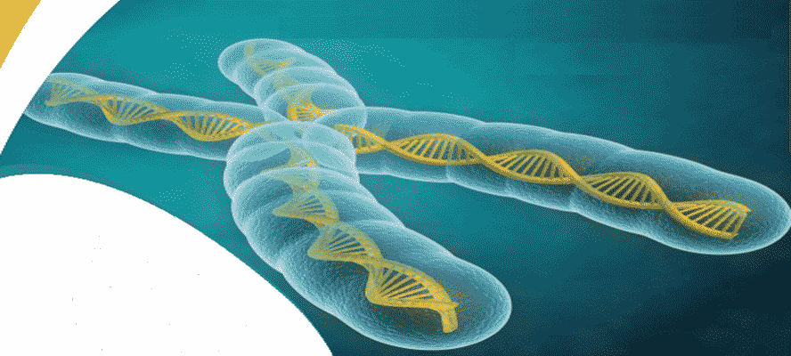
Genetic disorder (GD) is a condition resulting from an abnormality in an individual’s DNA, either a small
mutation in one gene or a change in the chromosome structure or number. There are around 4,000 to 6,000
Genetic disorders diagnosed till date. With the higher than ever birthrate, increasing incidences of genetic disorders are
observed in newborns in India, with half a million affected by malformations while 21,000 with Down syndrome.
Some Genetic disorders can be inherited from parents as a result of mutations present on either of the two copies of genes received by the offspring from the parents. The chances of acquiring this kind of disorders depends upon whether the genes are autosomal dominant, autosomal recessive or sex linked. These include Cystic Fibrosis, Tay Sachs, and Sickle Cell Disease.
Types of Genetic Disorders
Genetic disorder can be broadly classified into three categories
- Single Gene Disorders – Phenylketonuria Single gene or Mendelian disorders involves mutation that affect a single gene and includes autosomal dominant (AD) and recessive (AR), X-linked recessive (XR) and dominant (XD), and Y-linked disorders. It is the most prevalent inherited defect caused by error in amino acid metabolism. In this autosomal recessive disorder, mutation occurs in the gene encoding the enzyme phenylalanine hydroxylase which leads to failure in hydroxylation of phenylalanine to tyrosine. This results in increased concentration of phenylalanine in blood, which if left untreated can lead to severe mental retardation, epilepsy and behavioral problems. Accumulation of phenylketones is usually observed in the affected individuals.
- Chromosomal Disorders. Down syndrome. Chromosomal disorders comprises of excess or deficiency of genes, change in structure or number of chromosome. These disorders results from chromosomal aberrations which include numerical, occurring due to errors at chromosome pairing and, crossing over and structural damages. Trisomy of chromosome 21 is one the commonest human autosomal aneuploidy disorders affecting 1 in 800 to 1 in 1000 live born infants. Nondisjunction of the chromosome 21 during egg or sperm development is the reason for addition of an extra chromosome in the zygote. This condition is usually characterized by learning disabilities, mental retardation, craniofacial abnormality and hypotonia in early infancy while also associated with congenital heart defects, leukemia, and Alzheimer’s disease. Physical manifestation of this syndrome includes small chin, slanted eyes, flat nasal bridge, single crease of the palm, protruding face (due to small mouth and large tongue), big toe, abnormal pattern of fingerprint and short fingers.
- Complex Disorders – Coronary Artery Disease (CAD) Complex or Multifactorial disorders results from combination of mutations in two or more genes, usually influenced by environmental factors and lifestyle. Most types of cleft lip/palate, club foot, neural tube defects, Coronary Artery Disease, hypertension, colon cancer and diabetes are some of the common disorders that are enlisted as complex genetic disorders. Coronary Artery Disease is one of the global leading cause of mortality and results due to the atherosclerotic plaque that builds up in the blood vessels. Familial hypercholesterolemia, a hereditary pattern of increased cholesterol resulting from mutations in low density lipoprotein (LDL) receptor gene. Predisposing factors of Coronary Artery Disease includes age, gender (male), smoking, elevated blood pressure, diabetes, obesity and a sedentary lifestyle. While increased concentrations of circulating LDL cholesterol, triglyceride rich lipoproteins, or decreased high-density lipoprotein HDL cholesterol are also linked to risk for Coronary Artery Disease.
Types of Genetic Screening
Mostly classified as Premarital, Preconception, Prenatal and Newborn screening.
1. Premarital Screening
This screening works in identifying the at risk couples i.e. possibility of a couple being carrier of any contagious
disease, genetic defect or disorder. Consanguineous marriage is one of the main reasons for prevalence of genetic
defects in families. In India, incidences of such marriages are still prevalent and results in genetic transmission
of disease.
Screening for sickle cell, Beta-thalassaemia and G6PD deficiency along with testing for HIV, Rh factor, hepatitis, and others are performed by a blood tests. Following the tests, counseling can be undertaken, where the associated risks are explained to the couple and following steps can be considered
- Avoiding marriages with partners having higher carrier risk, or
- Avoiding having children and opting for artificial insemination by donor, egg donation or adoption.
Importance of Karyotyping as a part of Premarital Screening
Karyotyping can be beneficial in detection of following
- Turner’s syndrome
- Klinefelter’s syndrome
- Balanced translocations
- Polymorphic variations
Preconception Screening
Preconception screening can be done by couples before planning for conception to check for the possible
unknown threats such as infectious diseases like HPV, Hepatitis B, HIV or sexually transmitted diseases
carried by them. Preconception carrier screening can be useful to check couples for being potential carriers
of some disorders that may manifest in their child.
Newborn Screening (NBS)
History of NBS dates back to Dr. Asbj??rn F??lling, a biochemist from Norway who was the first person to
discover ‘Imbecillitas phenylpyruvica’ in 1934 which was later named as PKU or Phenylketouria. Further,
development of dried blood on filter paper test by Dr. Robert Guthrie in 1961 proved a life saving test for
the affected newborns to be provided with special diet to prevent the brain damage. This was followed by
gradual addition of different screening tests to the panel which helped in further diagnosis and giving a
window for early treatment that can be provided to the newborn. Incidences of birth defects accounts
approximately 6-7% in India. Congenital anomalies in India, are responsible for 815% of perinatal deaths
and 13-16% of neonatal deaths.
NBS also known as Neonatal Screening is usually done within few hours after the baby’s birth with a heel-stick technique which is collection of small amount of blood sample from the newborn’s heel. Newborns are screened for Amino Acid, Organic Acid and Fatty Acid Oxidation Disorders and other disorders like congenital hypothyroidism, PKU, sickle cell anemia, cystic fibrosis, SCID and other lysosomal storage disorders. It also includes tests for infections such as HIV and Hepatitis B. Few of these NBS are enlisted below
Cystic Fibrosis (CF)
CF is a disorder which involves abnormal cationic transport across cell membranes i.e. mutated gene encoding
a defective chloride channel in epithelial cells named the cystic fibrosis transmembrane conductance regulator
(CFTR). This causes highly viscous secretions, affecting many organ systems but primarily the gastrointestinal
tract and lungs. Infants with 1 or 2 detected mutations are referred for diagnostic sweat testing.
Congenital Hypothyroidism (CH)
Congenital Hypothyroidism is one of the common preventable cause of mental retardation. It results mostly due to abnormal development
of the thyroid gland (thyroid dysgenesis) and dyshormonogenesis. Detection of Congenital Hypothyroidism depends on the immunoassay
measurement of various combinations of TSH, thyroxine and thyroxine-binding globulin. Due to subtle clinical
manifestations it remains undiagnosed at birth and aggravating later as mental retardation. This emphasizes
the importance of neonatal screening, blood
spot TSH or thyroxine (T4) or both are carried out by heel prick test.
Medium-chain acyl-CoA dehydrogenase deficiency (MCAD)
MCAD is a fatty acid metabolism disorder which results due to the lack of one of the enzyme involved in
mitochondrial fatty acid B-oxidation. This enzyme is required to fuel hepatic ketogenesis, a major source
of energy when the hepatic glycogen stores becomes depleted. MCAD is screened by detecting elevated
blood levels of octanoylcarnitine.
Prenatal Screening and Diagnosis
Screening Tests
Prenatal or antenatal screening can be done by a pregnant women to screen for any genetic defect or
disease being carried by the fetus. These tests mostly include screening for aneuploidy and neural tube defects.
Incidence of chromosomal abnormalities mostly trisomy is affected by maternal age, chances increases with advancing age. Hence age is the main risk factor which demands prenatal screening. Other factors include family history of some genetic disorders, previous baby with birth defect or parents being carriers.
Ultrasound mostly forms the primary level of screening and is performed throughout the pregnancy, this is usually followed by a simple blood test of the mother. Ultrasound tests such as nuchal translucency can detect the fluid backup under the skin or the at the backside of developing neck which can be an indicative of trisomy 18, Down syndrome or even heart defect. Different blood tests performed during different trimesters such as
- First Trimester
Pregnancy Associated Plasma Protein A (PAPP-A) and serum free B-human chorionic gonadotropin
(free Beta hCG), are the protein and hormone respectively produced by the placenta. Decreased levels
of PAPP-A and elevated levels of free B-hCG are observed in Down syndrome while decreased B-hCG
is observed in trisomies of chromosome 18 and 13.
Non-Invasive Prenatal Genetic Testing (NIPT)- NIPT uses cell-free fetal DNA (cffDNA) in the blood of a pregnant woman for detection of fetal chromosomal aneuploidies. NIPT propounds as a mediator step between serum screening and invasive diagnostic testing for determination of any likelihood of fetal aneuploidy. It is usually carried out after 10 weeks of pregnancy and detects chromosomal aneuploidies like Down, Edward, Patau, Turner and Klinefelter Syndrome. - Second Trimester Inhibin A, a hormone produced by placenta and its abnormal high levels are seen in Down syndrome. Alpha-fetopotein (AFP) produced by fetus is passed into mother’s serum from where it can be detected. Elevated levels can indicate neural tube defects such as open spina bifidia, anencephaly as well as Edward and Down syndromes. Unconjugated estriol, produced by fetus in lower amount while elevated levels are indicative of trisomy of chromosome 18 and 21 i.e Edward and Down syndromes.
- Third Trimester Usually comprises urine analysis for protein, glucose and bacterial detection. CBC (complete blood count), estimation of thyroid hormone as well as detection of HIV, HBV, STDs and other infectious diseases.
Diagnostic Tests for Chromosomal Disorders
There are two main techniques used for collection of samples for prenatal diagnostic tests – Amniocentesis
and Chorionic Villus Sampling (CVS).
- Amniocentesis It is usually carried out between 15″ to 20″ week of pregnancy and involves extraction of small sample of amniotic fluid (it contains cells from the baby), with the help of a needle. It can be used to detect neural tube defects like spina bifida and chromosomal abnormalities (monosomy/trisomy).
- CVS It is usually carried out before the amniocentesis, between 10 to 12″ week of pregnancy. Small sample of fetal chorionic villi cells are extracted from the placenta.
Disadvantage of these diagnostic tests include risk of miscarriage in <1% population.
Sample collected either in the form of amniotic fluids or chorionic villi cells are further analyzed by Karyotyping, Fluorescence in situ hybridization (FISH) and Microarray.
- Karyotyping
Karyotyping is the process of chromosome identification by obtaining a karyotype that is a compendium of an individual’s chromosomes. A photographic presentation of these chromosomes is called a karyogram. Samples used for this process include samples from bone marrow, blood, placenta or amniotic fluid. For visualization of the chromosomes they are stained with a dye, this is called as chromosome banding. These bands help in identification and differentiation of the normal and abnormal chromosomes as well as information of various structural and numerical abberations. -
Fluorescence In situ Hybridization (FISH)
FISH is a rapid testing technique which utilizes fluorescence tagging of entire or portions of chromosomes and aids in their visualization. Numerical as well as structural chromosomal abnormalities such as aneuploidies and microdeletion can be detected by this technique. Advantages of FISH over karyotyping are, it is rapid and can be done on non-dividing cells, for analysis. Limitation of this technique is that it does not provide an account of complete set of 44 autosomes and a pair of sex chromosome. -
Chromosomal Microarray Analysis (CMA)
CMA also known as array-based copy-number analysis is used to identify point mutations, and numerical and structural chromosome abnormalities. CMA utilizes fluorescently labeled DNA that is hybridized to a slide containing thousands of probes which are spread across the genome. Intensity of fluorescence obtained from the binding of DNA and specific probes is measured. This helps in identification of regions with extra copies of DNA, indicated by higher fluorescence or regions with missing copies of DNA indicated by lower fluorescence. CMA has higher resolution as compared to conventional karyotyping as it identifies chromosomal abnormalities, including submicroscopic abnormalities.
Screening of disorder should be followed by a confirmatory diagnostic test in high risk or positive screen cases which can give a clear and sure indication of the condition. To continue the pregnancy or undergo abortion is the decision to be taken. If the choice is to continue with the pregnancy then steps should be taken to inform themselves about the disorders and special arrangements should be done. Neonatologists and Pediatric sub-specialists can be consulted.
Some decisions are painful yet crucial and at such times making a decision is more important irrespective of it being right or wrong, the results of screening might create anxiety and stress but making a decision is better than waiting for the consequences.
Protect yourself and the coming generations … with the advent of technologies for healthy existence!


