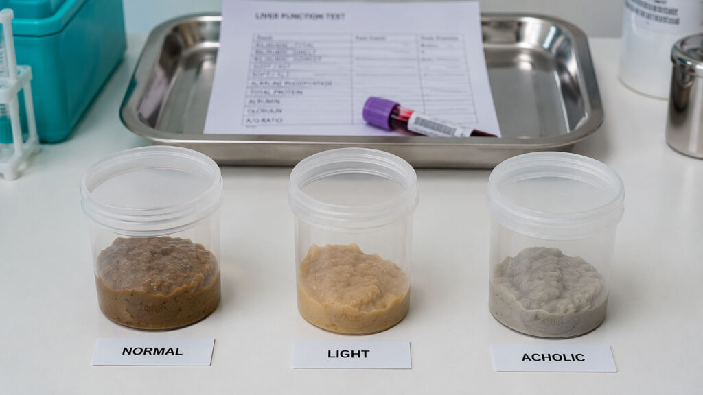
Most people learn something is wrong with their liver because of pain. A smaller group learns it because of a color. Bile is what makes stool brown, and when bile stops arriving, the change shows up in the toilet bowl days before anything starts to hurt. That head start is the whole value of the symptom, and the version that comes with no pain at all is often the one that matters most.
Table of Contents
Quick Answer: One pale stool is usually harmless. Pale or clay-colored stool lasting more than two days needs medical evaluation, and pale stool with dark urine, yellow eyes, itching, or right-sided pain needs it within 24 to 48 hours. The color means too little bile is reaching your intestine, either because the liver is not producing it or because a duct is blocked.
Stool color is one of the few internal readouts your body prints for free every day. Patients booking liver panels with us often describe the same sequence: the color shifted, they assumed it was something they ate, and only the third or fourth episode sent them looking for answers.
This guide covers what the color actually measures, how long is too long, the nine causes behind it, the harmless explanations worth ruling out first, and the exact point at which waiting stops being reasonable.
At a Glance
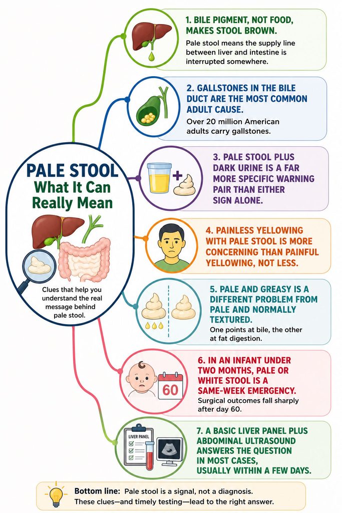
• Bile pigment, not food, makes stool brown. Pale stool means the supply line between liver and intestine is interrupted somewhere.
• Gallstones in the bile duct are the most common adult cause. Over 20 million American adults carry gallstones.
• Pale stool plus dark urine is a far more specific warning pair than either sign alone.
• Painless yellowing with pale stool is more concerning than painful yellowing, not less.
• Pale and greasy is a different problem from pale and normally textured. One points at bile, the other at fat digestion.
• In an infant under two months, pale or white stool is a same-week emergency. Surgical outcomes fall sharply after day 60.
• A basic liver panel plus abdominal ultrasound answers the question in most cases, usually within a few days.
How Long Is Too Long?
Duration is the variable readers ask about most, so here it is up front before anything else.
One episode, feeling fine: watch it. Diet, fast transit, and normal variation explain most single light stools. If the next two bowel movements are normal brown, you are done.
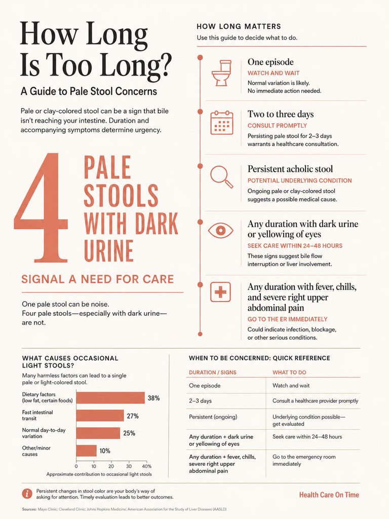
Two to three days of consistently pale stool: book an appointment. Major medical centers advise contacting a provider when light-colored stool persists beyond a few days, because persistent acholic stool usually reflects an underlying condition that worsens without care.
Any duration, with dark urine or yellow eyes: 24 to 48 hours. The pairing changes the math entirely.
Any duration, with fever, chills, and severe right upper abdominal pain: emergency room now.
Our medical reviewers use the same simple frame with patients: one pale stool is noise, four pale stools with dark urine is signal.
What Pale or Clay-Colored Stool Actually Means
How Bile Turns Stool Brown
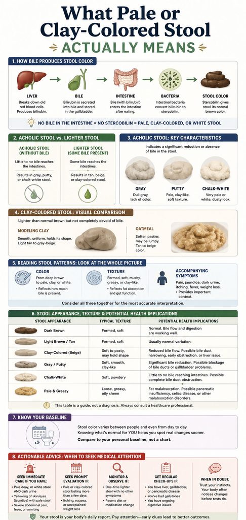
Your liver produces bile continuously. Bile carries bilirubin, the pigment left over when worn-out red blood cells break down, and drains through the bile ducts into the small intestine.
Once bilirubin reaches the colon, gut bacteria convert it into stercobilin. Stercobilin is the compound that makes normal stool brown. No stercobilin, no brown.
Stool color is therefore a daily report on bile flow. When it turns pale, one of two things has happened: the liver is not making enough bile, or something is blocking the pipe it travels through.
Acholic Stool Versus Merely Light Stool
Clinicians use the word “acholic,” meaning without bile, for stool that has lost its color entirely. That is the version signaling real biliary obstruction.
It is not the same as stool that is simply lighter than usual. Normal stool spans a wide band of brown and tan depending on transit time, diet, and hydration. A light tan stool after a heavy dairy weekend is not acholic.
The distinction changes the next step, which is why our medical team draws it carefully. True acholic stool is gray, putty, chalk-white, or the color of wet cement, and it holds that appearance across several bowel movements.
What Clay-Colored Looks Like in Real Life
People describe it consistently: modeling clay, unfinished pottery, oatmeal, drywall compound. Some describe beige with a faint greenish cast.
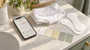
Your own baseline is the most reliable comparison. If you look twice and think “that is not my usual color,” photograph it. Bathroom lighting distorts color badly, and a daylight photo gives your doctor something objective to work from.
Texture matters as much as color, which is where most online guides go wrong.
Table 1: Reading Your Own Stool Pattern
| Appearance | Texture and float | Common companion signs | Most likely cause group | Urgency window |
| Gray, putty, or clay; normal formed texture | Normal, usually sinks | Dark urine, yellow eyes, itching | Bile duct obstruction or liver disease | 24 to 48 hours |
| Pale, bulky, greasy, foul-smelling | Loose, floats, leaves an oil film | Weight loss, bloating, gas, cramping | Fat malabsorption (pancreas or small intestine) | 1 to 2 weeks; sooner with weight loss |
| Chalky white, one to three days | Normal | Recent barium X-ray or CT | Barium sulfate contrast passing through | No action needed |
| Pale or white in infant under 8 weeks | Variable | Persistent jaundice, dark urine, poor weight gain | Biliary atresia until proven otherwise | Same week, ideally same day |
| Clay-colored with fever, chills, severe right upper pain | Any | Confusion, low blood pressure | Ascending cholangitis (infected blocked duct) | Emergency room immediately |
| Light tan, intermittent, resolves within a day | Normal | None | Diet, transit speed, normal variation | Monitor only |
The same color carries six different meanings depending on what surrounds it. This is precisely why “pale stool” alone is a weak signal and “pale stool plus X” is a strong one.
The 9 Causes of Pale and Clay-Colored Stool
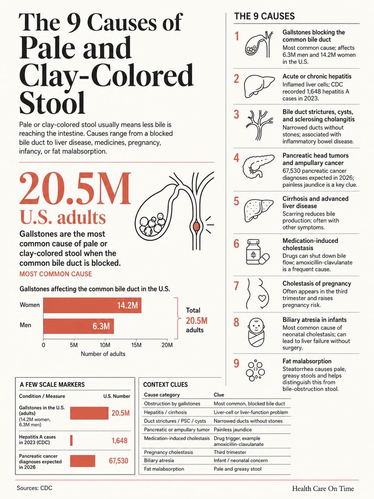
1. Gallstones Blocking the Common Bile Duct
This is the most common adult cause in the United States by a wide margin. A stone formed in the gallbladder migrates into the common bile duct and lodges there, damming bile upstream.
Gallstones are extraordinarily common. Over 6.3 million men and 14.2 million women aged 20 to 74 in the US have them, and prevalence climbs with age, exceeding 25% in women past 60. Most carriers never know.
Why Most Gallstone Carriers Never See This Symptom
Roughly 10% of people with gallstones develop symptoms within 5 years and 20% within 20 years. Having stones is not the risk. Having one migrate into the duct is.
When it does, the classic pattern is right upper abdominal pain arriving in waves, often after a fatty meal, followed within a day or two by dark urine and pale stool. Some stones pass on their own and color returns to normal. Some do not.
Patients often ask us whether gallbladder pain is always severe. It is not. Duct stones can produce a dull ache or nothing at all, particularly in older adults and people with diabetes.
2. Acute or Chronic Hepatitis
Inflamed liver cells cannot process and secrete bile normally. Viral hepatitis A, B, and C are the most common infectious causes, and alcohol-associated and toxic hepatitis produce the same result.
During 2023, CDC recorded 1,648 reported hepatitis A cases nationally, corresponding to an estimated 3,300 actual infections after adjusting for underreporting, a 28% decline from 2022. The chronic viruses carry the heavier load: an estimated 660,000 US adults had hepatitis B and 2.2 million had hepatitis C across the 2017 to 2020 period.
Hepatitis-related pale stool usually arrives alongside fatigue, appetite loss, nausea, and low-grade fever, often several days before visible yellowing.
3. Bile Duct Strictures, Cysts, and Sclerosing Cholangitis
Ducts narrow without a stone involved. Scarring from previous surgery, chronic inflammation, or autoimmune attack all produce strictures.
Primary sclerosing cholangitis progressively scars the bile ducts and carries a strong association with inflammatory bowel disease. It typically produces fluctuating symptoms over months rather than an abrupt change.
Choledochal cysts, congenital dilations of the bile duct, can surface in adulthood with the same picture.
4. Pancreatic Head Tumors and Ampullary Cancer
The common bile duct passes directly through the head of the pancreas on its way to the intestine. A tumor there compresses the duct from the outside.
An estimated 67,530 Americans will be diagnosed with pancreatic cancer in 2026, and more than 52,740 will die from it. The all-stage 5-year relative survival rate is 13%, and median age at diagnosis is 71.
Painless Jaundice Outranks Painful Jaundice
This detail gets buried in most consumer health content. Painless obstructive jaundice is the most characteristic sign of cancer in the head of the pancreas, and patients typically notice darkening urine and lightening stool before anyone notices their skin has changed.
Clinicians can usually detect jaundice once total bilirubin reaches 2.5 to 3 mg/dL. Patients and families usually do not notice it until bilirubin reaches 6 to 8 mg/dL. That gap is measured in weeks, and it is exactly why the stool color change earns its place as an early signal.
None of this means pale stool means cancer. Pancreatic cancer is not the most common cause of jaundice; gallstones, hepatitis, and other liver and bile duct conditions are far more common. It means pale stool without pain deserves the same urgency as pale stool with pain, not less.
5. Cirrhosis and Advanced Liver Disease
Extensive scarring reduces the liver’s capacity to produce bile at all. Cirrhosis from any cause, including steatotic (fatty) liver disease, alcohol, and chronic viral hepatitis, can produce persistently pale stool in later stages.
Here the stool change rarely arrives alone. Swelling in the legs and abdomen, easy bruising, spider-like vessels across the chest, and mental fogginess usually accompany it.
6. Medication-Induced Cholestasis
Drugs can shut down bile flow at the cellular level with no physical blockage anywhere. Amoxicillin-clavulanate, sold as Augmentin, is the single most frequent cause of idiosyncratic drug-induced liver injury in the US Drug-Induced Liver Injury Network registry, accounting for 11% of all cases and 24% of antimicrobial cases.
The timing catches people out. Onset runs from a few days to 8 weeks after starting the drug, averaging about 3 weeks, and it frequently begins after the antibiotic course has already finished. Few patients connect a completed prescription to a new symptom.
The injury pattern is predominantly cholestatic, with sharply elevated alkaline phosphatase. The usual sequence is fatigue and nausea, then itching, then jaundice and pale stool.
Other agents linked to cholestatic injury include anabolic steroids, some oral contraceptives, azathioprine, and certain antipsychotics. Never stop a prescribed medication on your own; call the prescriber the same day instead.
7. Cholestasis of Pregnancy
Intrahepatic cholestasis of pregnancy typically appears in the third trimester. Its hallmark is intense itching, often worst on the palms and soles and worst at night, and it can precede any color change.
Pale stool and dark urine may follow. The condition carries real risks for the pregnancy, so any pregnant reader with unexplained itching should contact their obstetric provider rather than waiting for a stool change to confirm anything.
8. Biliary Atresia in Infants
In newborns, the bile ducts outside the liver can be progressively destroyed by inflammation and scarring. Without surgery this leads to liver failure within roughly a year.
Biliary atresia is the most common cause of neonatal cholestasis and accounts for nearly half of all pediatric liver transplants in the United States. Affected infants develop jaundice and pale, acholic stools within the first weeks after birth.
The pediatric section below covers the timing in full, because the window is narrow and the numbers are unforgiving.
9. Fat Malabsorption, the Non-Biliary Cause
Almost no consumer article separates this out, and separating it out changes the entire workup.
Steatorrhea is excess fat in the stool. It produces pale, large-volume, foul-smelling, loose stools that float and leave an oily film. The paleness comes from fat content, not from missing bile pigment.
Its causes sit in a different anatomical neighborhood: exocrine pancreatic insufficiency from chronic pancreatitis or cystic fibrosis, celiac disease, tropical sprue, giardiasis, small intestinal bacterial overgrowth, and Crohn’s disease. Fat-digesting enzyme output generally has to fall to roughly 5% to 10% of normal before steatorrhea appears.
The practical distinction is simple. Bile-obstruction stool is pale with normal texture and comes with dark urine and yellow eyes. Malabsorption stool is pale, greasy, and floats, and comes with bloating, gas, and gradual weight loss. Our lab partners see two entirely different panels ordered for these two pictures, which is why describing texture accurately to your doctor saves real time and money.
The Harmless Causes People Panic About
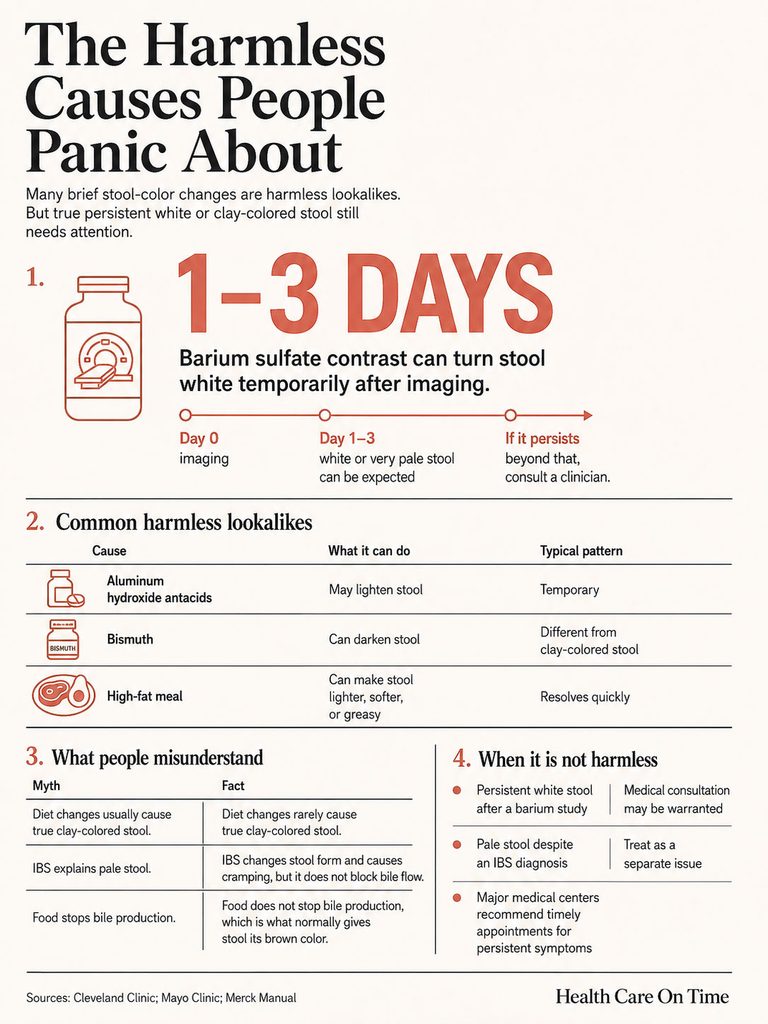
Barium After an Imaging Study
Barium sulfate contrast used in upper GI series and some CT protocols passes through undigested. It reliably turns stool chalky white or very light for one to three days afterward.
This is expected and needs nothing beyond normal hydration. White stool persisting well beyond several days after a barium study is worth a call.
Antacids, Anti-Diarrheals, and Very High-Fat Meals
Large doses of aluminum hydroxide antacids can lighten stool. Bismuth-containing products more often darken it, sometimes dramatically, which surprises people expecting the opposite.
A single very high-fat meal can speed transit and produce one lighter, softer stool. It resolves with the next meal.
Why Diet Almost Never Causes True Clay-Colored Stool
Food changes stool color at the margins. Beets redden it, leafy greens can green it, heavy dairy can lighten it a shade.
Food does not produce gray, putty, or chalk-white stool, because food does not stop bile production. Any explanation requiring you to have eaten something unusual for three consecutive days is usually the wrong explanation.
What IBS Does and Does Not Do
Irritable bowel syndrome causes cramping, bloating, urgency, and alternating constipation and diarrhea. It does not block bile flow and it does not damage liver tissue.
IBS does not produce clay-colored stool. If you carry an IBS diagnosis and your stool turns pale, treat that as a new and separate problem, not a flare.
Symptoms That Change the Urgency
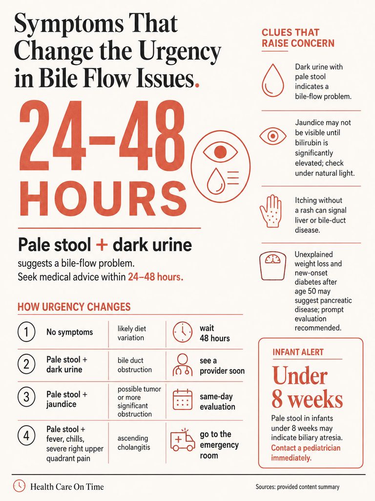
Dark Urine Plus Pale Stool
This pairing is the most useful clue on the page. When bile cannot exit through the intestine, conjugated bilirubin backs up into the bloodstream and the kidneys clear it into urine.
Stool loses pigment at the same moment urine gains it. Tea-colored or cola-colored urine alongside light stool is close to diagnostic for a bile flow problem and should not be watched for a week.
Either sign alone can be innocent. Dehydration darkens urine. A single light stool means little. Together they mean something.
Jaundice and Why Patients Notice It Late
Yellowing appears in the whites of the eyes before the skin, and it shows earlier in natural daylight than under indoor lighting. Check in front of a window, not a bathroom mirror.
Because visible jaundice lags the underlying bilirubin rise by days, the absence of yellow eyes does not rule out obstruction.
Itching Without a Rash
Bile salts accumulating under the skin cause pruritus, and it often precedes jaundice. Patients frequently rate it as their most distressing symptom.
Generalized itching with no visible rash, worst at night and on palms and soles, is a liver and bile duct symptom until proven otherwise.
Fever, Chills, and Right Upper Pain
Fever plus jaundice plus right upper quadrant pain is Charcot’s triad, pointing to ascending cholangitis, an infection of an obstructed bile duct. Add confusion and low blood pressure and it becomes Reynolds’ pentad.
This is an emergency department presentation, not a next-day appointment. Blocked, infected bile becomes bloodstream infection quickly.
Unexplained Weight Loss and New-Onset Diabetes
Losing weight without trying, alongside pale stool, shifts the differential toward malignancy and chronic pancreatic disease. New-onset type 2 diabetes in an adult over 50 with no risk factors, particularly with weight loss, is a recognized red flag for pancreatic disease.
Table 3: Scenario to Action
| Your scenario | What it most likely means | What to do, and how fast |
| One pale stool, feel fine, no other symptoms | Diet, transit speed, or normal variation | Monitor 48 hours. No appointment needed if it normalizes |
| Chalky stool 1 to 3 days after a barium X-ray or CT | Contrast agent passing through | Nothing. Hydrate normally |
| Pale stool 3+ days, no pain, no other symptoms | Early cholestasis or slow-growing obstruction | Liver panel and provider visit within 3 to 5 days |
| Pale stool plus dark urine, with or without pain | Bile duct obstruction or acute liver injury | Provider within 24 to 48 hours; sooner if yellowing appears |
| Pale stool plus yellow eyes, no pain, unexplained weight loss | Possible tumor compressing the bile duct | Same-day or next-day appointment. Do not wait it out |
| Pale stool plus fever, chills, severe right upper pain | Ascending cholangitis | Emergency room immediately |
| Pale, greasy, floating stool plus weight loss over weeks | Fat malabsorption, pancreatic or intestinal | Provider within 1 to 2 weeks; sooner with rapid weight loss |
| White or pale stool in an infant under 8 weeks | Biliary atresia until proven otherwise | Call the pediatrician today and request a direct bilirubin |
Pale Stool in Babies and Children
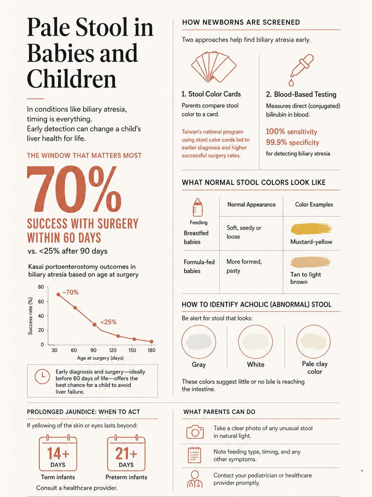
The 60-Day Window That Decides Outcomes
Biliary atresia is treated with the Kasai portoenterostomy, a surgery reconstructing bile drainage. Timing determines whether it works.
If the Kasai is performed within the first 60 days of life, roughly 70% of infants establish bile flow. Beyond 90 days, fewer than 25% do.
That is the entire reason infant stool color is treated so seriously. There is no way to buy the time back.
The difficulty is that jaundice is extremely common and mostly benign in newborns, and infants with biliary atresia often look well early on. The disease hides inside a normal-looking baby.
Stool Color Cards and the Two-Week Bilirubin Rule
Two screening approaches have been studied. The first is the infant stool color card, a printed reference strip given to new parents showing normal and abnormal stool colors side by side. Taiwan’s national card program produced earlier diagnosis, earlier Kasai surgery, and improved jaundice-free survival at three years.
The second is blood-based. A study of 124,385 newborns published in JAMA in 2020 tested a two-stage screen using direct or conjugated bilirubin: all newborns tested within the first 60 hours of life, with those screening positive retested at or before the two-week visit. The approach identified all 7 known biliary atresia infants, with 100.0% sensitivity and 99.9% specificity, though the confidence interval around sensitivity was wide.
Current North American and European pediatric gastroenterology guidance recommends that any infant still jaundiced at 2 weeks of age be evaluated for cholestasis with total and direct bilirubin measurement. Prolonged jaundice is defined as jaundice continuing past 14 days in term infants and past 21 days in preterm infants.
If your two-week-old is still yellow, that measurement is the ask. Parents who bring us questions about newborn testing are often surprised how simple and inexpensive the first test is.
Normal Newborn Stool Colors Parents Mistake for Pale
Breastfed infants pass mustard-yellow, seedy stool that can look startlingly light. Formula-fed infants pass tan to light brown stool. Meconium in the first days is near-black and tarry.
None of those are acholic. Acholic infant stool is gray, white, or pale clay, with no yellow or green cast at all. Photograph it in daylight and bring the photo to the appointment.
Brightly colored foods explain most odd stool colors in older children. They do not explain white or gray.
What the Numbers Say About the Conditions Behind Pale Stool
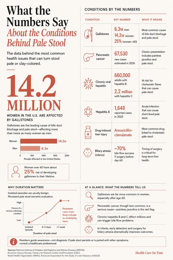
Table 2: US Data on the Conditions That Cause Pale Stool
| Condition | US figure | Why it matters for pale stool | Source |
| Gallstones | 6.3 million men and 14.2 million women aged 20 to 74 affected; over 25% of women past 60 | Largest reservoir of adults at risk of a duct-blocking stone | StatPearls, Cholelithiasis (NBK459370) |
| Symptomatic progression | About 10% symptomatic within 5 years, 20% within 20 years | Most stone carriers never develop pale stool | StatPearls, Cholelithiasis |
| Pancreatic cancer | 67,530 estimated new US cases and 52,740 deaths in 2026; 13% five-year survival | Painless jaundice with pale stool is the classic head-of-pancreas presentation | American Cancer Society, Cancer Facts & Figures 2026 |
| Chronic viral hepatitis | An estimated 660,000 adults with hepatitis B and 2.2 million with hepatitis C, 2017 to 2020 | Large population at risk of cholestatic flares | CDC, Viral Hepatitis Surveillance |
| Hepatitis A | 1,648 reported cases in 2023, an estimated 3,300 infections, down 28% from 2022 | Acute cause of short-lived pale stool with jaundice | CDC, 2023 Viral Hepatitis Surveillance Report |
| Drug-induced liver injury | Amoxicillin-clavulanate is 11% of all DILIN cases and 24% of antimicrobial cases | Most common single drug behind cholestatic pale stool | Drug-Induced Liver Injury Network registry |
| Biliary atresia surgery timing | About 70% establish bile flow if Kasai occurs before day 60; under 25% after day 90 | Defines the infant emergency window | Pediatric hepatology literature, NASPGHAN guidance |
How Often Pale Stool Turns Out to Be Serious
There is no clean national statistic on what share of pale stool episodes prove dangerous, and any page quoting one is inventing it. What the evidence does support is a pattern.
Isolated single episodes with no accompanying symptoms are usually benign. Persistent acholic stool, meaning several days running, usually reflects an underlying condition that worsens without care.
Duration and companion symptoms carry the information. That is the honest answer, and it is more useful than a fabricated percentage.
How Doctors Find the Cause
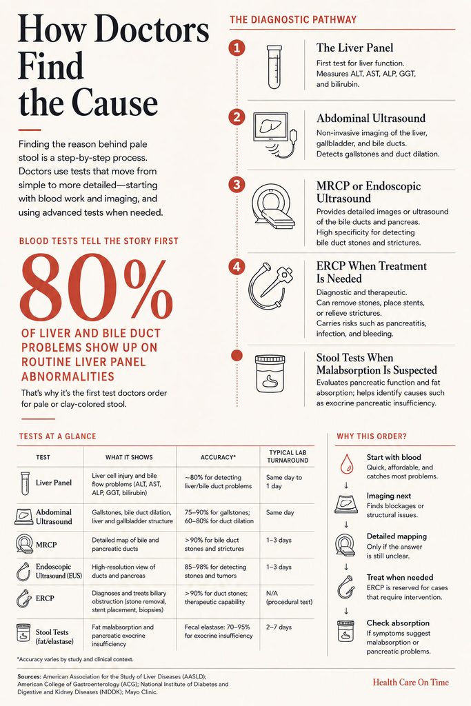
Step 1: The Liver Panel
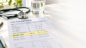
Nearly always the first test, and it is inexpensive and fast. It measures alanine aminotransferase (ALT), aspartate aminotransferase (AST), alkaline phosphatase (ALP), gamma-glutamyl transferase (GGT), and total plus direct bilirubin.
The pattern matters more than any single value. Sharply elevated ALP and GGT with high direct bilirubin points at obstruction. Sharply elevated ALT and AST points at liver cell injury such as hepatitis or drug toxicity.
Readers wanting the deeper breakdown can review our detailed guides on elevated ALT and elevated AST, which cover reference ranges and the causes behind each pattern.
Step 2: Abdominal Ultrasound
Ultrasound is first-line imaging. It is painless, radiation-free, and good at spotting gallstones, a dilated bile duct, and gross liver abnormalities.
A common bile duct wider than about 6 mm with the gallbladder still in place is considered dilated and suggests obstruction downstream.
Ultrasound has a known weakness: it struggles to see the lower end of the bile duct because overlying bowel gas obscures it. A normal ultrasound does not close the case, and knowing that is what keeps a workup from stopping too early.
Step 3: MRCP or Endoscopic Ultrasound
When ultrasound is inconclusive, the next tier is magnetic resonance cholangiopancreatography (MRCP) or endoscopic ultrasound (EUS). Both map the ducts in detail without treating anything.
Both carry high specificity for bile duct stones, reported at approximately 0.97 for EUS and 0.92 for MRCP, with EUS possibly more sensitive for small stones under about 6 mm. Current ASGE guidance suggests either test for intermediate-risk patients, with the choice driven by local expertise, availability, and patient factors such as claustrophobia or a pacemaker.
Step 4: ERCP When Treatment Is Needed
Endoscopic retrograde cholangiopancreatography (ERCP) is both diagnostic and therapeutic. A scope passed through the mouth reaches the duct opening, and the endoscopist can remove stones, cut the sphincter, or place a stent in the same session.
ERCP is minimally invasive but not risk-free, with adverse events in 6% to 15% of patients, pancreatitis being the most common. Guidelines therefore reserve it for people likely to need treatment rather than using it to look around.
ASGE high-risk criteria justifying direct ERCP are: a bile duct stone visible on ultrasound or cross-sectional imaging, or total bilirubin above 4 mg/dL combined with a dilated common bile duct, or ascending cholangitis.
Stool Tests When Malabsorption Is Suspected
If the picture is greasy floating stool rather than clay-colored formed stool, the tests change entirely. Fecal elastase measures pancreatic enzyme output. Quantitative fecal fat over a defined collection period confirms steatorrhea.
Celiac serology, stool studies for Giardia and other parasites, and breath testing for bacterial overgrowth complete the panel. In cases reviewed by our medical team, the most frequent avoidable delay is ordering a bile-duct workup for what was always a pancreatic enzyme problem.
Table 4: What Each Test Shows and How Fast
| Test | What it answers | Accuracy or key threshold | Typical turnaround |
| Liver panel (ALT, AST, ALP, GGT, bilirubin) | Is this obstruction, liver cell injury, or neither | ALP and GGT dominant pattern suggests cholestasis | Same day to 24 hours |
| Abdominal ultrasound | Gallstones, duct dilation, liver texture | Duct above 6 mm with gallbladder present is dilated | Same day; report in 24 hours |
| MRCP | Detailed map of ducts without a scope | Specificity around 0.92 for duct stones | 1 to 3 days for scheduling and read |
| Endoscopic ultrasound (EUS) | Small stones and pancreatic head detail | Specificity around 0.97; better for stones under 6 mm | Often 1 to 2 weeks to schedule |
| ERCP | Confirms and treats in one session | Adverse events in 6% to 15% | Same-day when urgent |
| Fecal elastase | Pancreatic enzyme output when stool is greasy | Low result indicates exocrine insufficiency | 3 to 7 days |
Cost, Access, and What to Expect in the US
A basic liver panel is one of the least expensive tests in American medicine and is widely available through direct-access lab services without a physician order in most states. Cash prices commonly run in the low tens of dollars.
Abdominal ultrasound is the more variable line item, and it usually needs an order. Costs differ substantially between hospital outpatient departments and independent imaging centers, so it is worth asking for the CPT code and calling ahead if you are paying out of pocket.
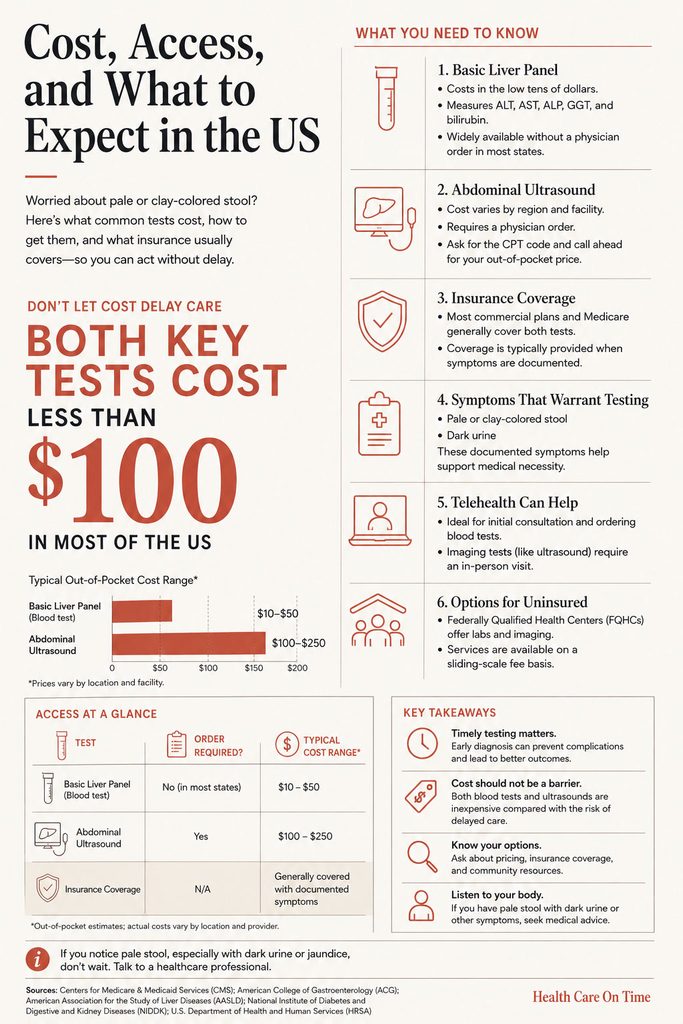
Most commercial plans and Medicare cover both tests when there is a documented symptom, and pale stool with dark urine is a documented symptom. Telehealth is reasonable for the first conversation and for getting the panel ordered, though imaging still requires an in-person visit.
If you are uninsured, federally qualified health centers use sliding-scale fees and can order both the panel and the ultrasound. Do not let cost push you into waiting weeks with acholic stool; the first two tests are the cheap ones.
Treatment by Cause
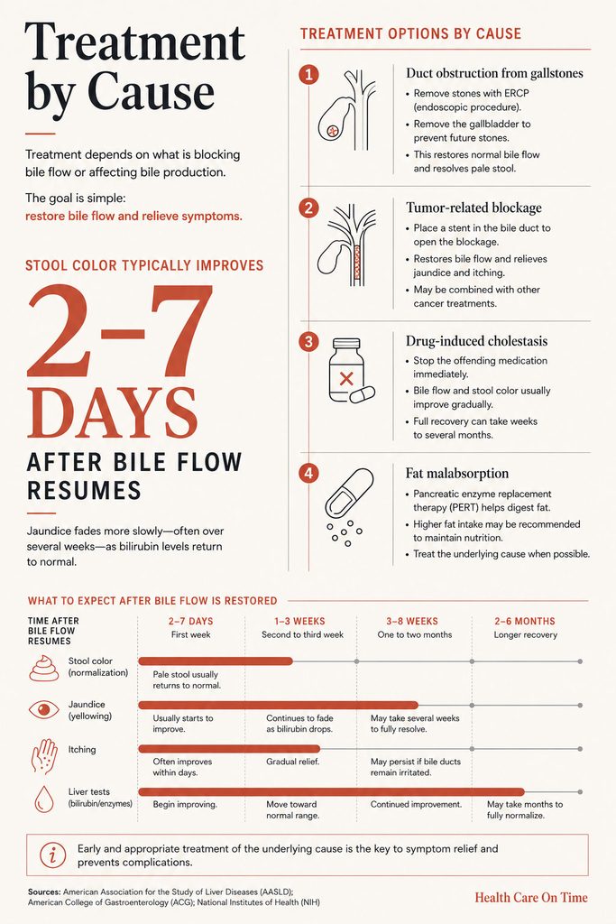
Stone Removal and Gallbladder Surgery
A stone in the duct is usually removed endoscopically during ERCP. The gallbladder is then typically removed, often laparoscopically, to prevent recurrence.
Stool color generally returns to normal within days of clearing the obstruction.
Stenting for Tumor-Related Blockage
When a tumor compresses the duct, a plastic or metal stent placed during ERCP restores drainage. This relieves jaundice and itching whether or not the tumor itself is treatable.
Restoring bile flow also improves nutritional status ahead of any surgery or chemotherapy.
Stopping the Offending Drug
For drug-induced cholestasis, removing the drug is the treatment. Recovery is often slow, sometimes taking months, though the eventual outcome for amoxicillin-clavulanate injury is usually full recovery.
Do not stop any prescription unilaterally. Call the prescriber, report the symptom, and let them handle the substitution.
Enzyme Replacement for Pancreatic Insufficiency
Pancreatic enzyme replacement therapy taken with meals restores fat digestion. Older advice to restrict dietary fat has been reversed, because fat restriction traded smaller stools for malnutrition.
Current practice is normal to higher fat intake paired with adequate enzyme dosing, plus monitoring of fat-soluble vitamins A, D, E, and K.
What Recovery of Stool Color Looks Like
Once bile flow resumes, color typically returns over two to seven days, often passing through a yellow-green phase before settling back to brown.
Jaundice fades more slowly than stool color normalizes, sometimes over several weeks, because pigment already deposited in tissue takes time to clear. A yellow tinge lingering after your stool is brown again is usually expected, not treatment failure.
What You Can Do Right Now
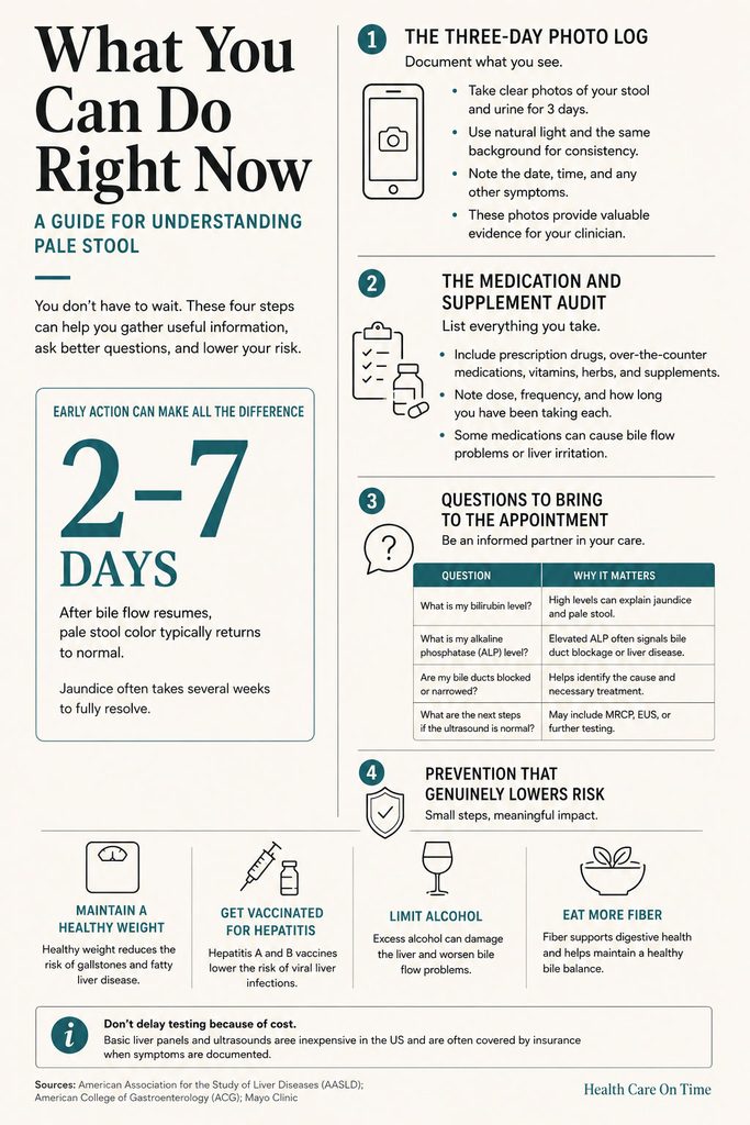
The Three-Day Photo Log
Photograph each abnormal stool in daylight and note the date, time, and any symptoms that day. Add a photo of your urine in a clear container against a white background.
This converts a vague complaint into evidence. It also settles the question of whether the color is genuinely abnormal or merely unusual for you.
The Medication and Supplement Audit
Write out every prescription, over-the-counter product, herbal supplement, and protein powder taken in the last three months, with approximate start dates.
Include courses you already finished. Cholestatic drug injury often begins after the bottle is empty, and this list is frequently the fastest route to an answer.
Questions to Bring to the Appointment
Ask directly: is my bilirubin mostly direct or indirect, is my alkaline phosphatase disproportionately high, is my bile duct dilated on imaging, and what is the plan if the ultrasound is normal but symptoms continue.
That last question matters most, because a normal ultrasound is exactly where many workups incorrectly stop.
Prevention That Genuinely Lowers Risk
Keeping weight in a healthy range lowers gallstone and chronic liver disease risk, though rapid crash weight loss raises gallstone risk sharply. Gradual loss is the safer route.
Hepatitis A and B vaccines exist and work well. Limiting alcohol protects the liver directly. Fiber intake, regular physical activity, and avoiding very long fasting periods all reduce gallstone formation.
None of this reverses an existing blockage. Prevention lowers the odds; it does not substitute for evaluation once the color has already changed.
Frequently Asked Questions
Is one pale stool a reason to worry?
Usually not. A single lighter-than-normal stool with no other symptoms is commonly explained by diet, fast transit, or normal variation. The threshold for concern is persistence. If stool stays pale across several bowel movements over two or more days, or if dark urine or yellow eyes appear, get evaluated.
How many days of clay-colored stool is too many?
Two days is a reasonable trigger for booking an appointment, and one day is enough if dark urine, jaundice, itching, or right-sided pain is present. Guidance from major medical centers advises contacting a provider for light-colored stool lasting more than a few days. Waiting a full week is not advisable.
Should I go to the ER for pale stool?
Pale stool alone is not an emergency. Pale stool with fever, chills, and severe right upper abdominal pain is, because that combination suggests an infected blocked bile duct. Also go immediately for confusion, vomiting that prevents keeping fluids down, or rapidly deepening yellowing of the eyes and skin.
Can stress or anxiety cause pale stool?
No. Stress genuinely affects bowel habits, speeding transit and altering consistency, and it can lighten stool by a shade. It cannot stop bile production or block a bile duct, so it does not cause true gray or clay-colored stool. Attributing acholic stool to stress delays diagnosis.
Does pale stool always mean liver cancer?
No, and cancer is far from the most common explanation. Gallstones, hepatitis, and medication effects account for the large majority of cases. That said, painless jaundice with pale stool in an adult over 50, particularly with weight loss, warrants prompt evaluation rather than watchful waiting.
Can IBS cause clay-colored stool?
No. Irritable bowel syndrome causes cramping, bloating, urgency, and swings between constipation and diarrhea, but it does not interfere with bile flow or damage liver tissue. If you have IBS and your stool turns clay-colored, treat it as a new problem needing its own workup, not as a flare.
Why is my poop pale after gallbladder removal?
Without a gallbladder, bile drips continuously into the intestine rather than releasing in concentrated pulses, which can lighten and loosen stool for weeks to months. This usually settles. Persistent clay-colored stool after surgery is different and can indicate a retained stone or bile duct injury, so report it.
Can antibiotics turn stool light-colored?
Yes. Amoxicillin-clavulanate is the leading single drug cause of idiosyncratic liver injury in US registry data and typically produces a cholestatic pattern. Onset averages about three weeks after starting and often begins after the course finishes. Call the prescriber rather than stopping medications on your own.
What does pale stool with dark urine mean?
It means bile pigment is not reaching your intestine and is being cleared by your kidneys instead. This pairing is far more specific for bile duct obstruction or acute liver injury than either sign alone and should prompt a liver panel and abdominal ultrasound within 24 to 48 hours.
Is white poop in a baby always serious?
Treat it as serious until a doctor says otherwise. White, gray, or pale clay stool in an infant under two months can indicate biliary atresia, where surgical outcomes fall sharply after day 60. Breastfed babies pass light mustard-yellow stool, which is normal; truly colorless stool is not.
Can a high-fat diet cause pale stool?
A single very fatty meal can produce one lighter, looser stool that resolves quickly. Persistent pale, greasy, floating, foul-smelling stool is different and points to fat malabsorption from pancreatic or small intestinal disease rather than from the meal itself, especially when weight loss accompanies it.
Which blood test detects a blocked bile duct?
The liver panel is the starting point, with alkaline phosphatase, GGT, and direct (conjugated) bilirubin carrying the most information about obstruction. Blood tests suggest the pattern but cannot locate the blockage, so abdominal ultrasound is ordered alongside them, with MRCP or endoscopic ultrasound if results stay unclear.
Medical Disclaimer: This article is provided by HealthCareOnTime for general information and education. It is not a substitute for professional medical advice, diagnosis, or treatment. Stool color changes can reflect conditions ranging from harmless to life-threatening, and only a qualified healthcare provider who has examined you and reviewed your results can determine the cause in your case. Never delay seeking medical care, and never start or stop any medication, because of something you read here. If you have pale stool with fever, severe abdominal pain, confusion, or rapidly worsening jaundice, seek emergency care immediately.
References
- Cleveland Clinic, Clay Colored Stool (Pale Stool)
- StatPearls, Cholelithiasis (NBK459370), National Library of Medicine
- StatPearls, Steatorrhea (NBK541055), National Library of Medicine
- LiverTox, Amoxicillin-Clavulanate (NBK548517), NIDDK
- Harpavat S, et al. Diagnostic Yield of Newborn Screening for Biliary Atresia Using Direct or Conjugated Bilirubin Measurements. JAMA. 2020;323(12):1141-1150
- ASGE Guideline on the Role of Endoscopy in the Evaluation and Management of Choledocholithiasis
- CDC, 2023 Viral Hepatitis Surveillance Report
- American Cancer Society, Signs and Symptoms of Pancreatic Cancer
- American Cancer Society, Key Statistics for Pancreatic Cancer
- NIDDK, Biliary Atresia
- Children’s Hospital of Philadelphia, Neonatal Jaundice: When to Refer
- deLemos AS, et al. Amoxicillin-Clavulanate-Induced Liver Injury. Dig Dis Sci. 2016