
Of every value printed on a standard blood panel, mean corpuscular volume is the one your body guards most tightly. National survey data suggests it fluctuates less within a single person than any other complete blood count measurement. So when your MCV moves, the movement deserves an explanation.
Table of Contents
Quick Answer
The MCV blood test measures the average size of your red blood cells, reported in femtoliters (fL). Most adult labs use a normal range near 80 to 100 fL. Below 80 fL is microcytic (small cells), usually iron deficiency. Above 100 fL is macrocytic (large cells), usually vitamin B12 or folate deficiency, alcohol, or medication. One abnormal MCV blood test result is a clue, not a diagnosis.
At a Glance
- Normal adult MCV runs roughly 80 to 100 fL, though some US labs print 79 to 97 fL instead
- Low MCV points toward iron deficiency or an inherited condition such as thalassemia trait
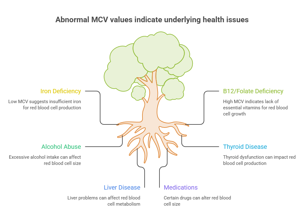
- High MCV points toward B12 or folate deficiency, alcohol, thyroid disease, liver disease, or medication
- RDW catches the deficiencies that MCV averages away, so a normal MCV does not clear you
- Red blood cells live about 120 days, so red blood cell size responds to treatment over months
- Two labs can report different MCV values for you in the same week, and both can be correct
- The large majority of abnormal MCV results trace back to treatable, everyday causes
What Your MCV Number Actually Measures
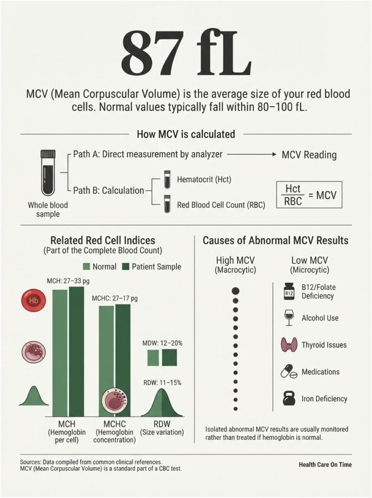
MCV stands for mean corpuscular volume. It is the average physical size of the red blood cells circulating in your sample at the moment blood was drawn.
Red blood cells exist to move oxygen from your lungs to your tissues. Their size records how well your bone marrow was supplied with raw materials while it was building them. Red blood cell size is a manufacturing log.
Femtoliters, and Why the Scale Is So Small
MCV is reported in femtoliters. A femtoliter is one quadrillionth of a liter, which is why results land in the 80s and 90s rather than in fractions.
At that scale, a shift of 4 or 5 fL is not rounding noise. It represents a real change in how your marrow assembles cells.
How Labs Produce the Number
Two methods exist. Modern hematology analyzers size each red cell directly as it crosses a sensor, then average thousands of individual measurements. Older or backup approaches calculate MCV by dividing hematocrit by red blood cell count and multiplying by ten.
The classic reference for red cell indices lists a normal value of 87 plus or minus 7 fL, with most clinical laboratories now using automated instruments built on either electrical impedance or optical detection. Platform choice matters, because different analyzers handle unusual cells differently.
Where MCV Sits Among the Red Cell Indices
MCV never travels alone. It belongs to a family of values calculated on every complete blood count, and reading it in isolation is the most common mistake patients make.
MCH describes the average hemoglobin per cell. MCHC describes hemoglobin concentration inside each cell. RDW describes how much the sizes vary from one cell to the next.
MCV vs MCH vs MCHC vs RDW in One Pass
Picture a bag of marbles. MCV is the average marble size. MCH is average weight. MCHC is density. RDW tells you whether the marbles all match or come in wildly mixed sizes.
Our medical reviewers see the same pattern repeatedly. Patients focus on the MCV blood test result because it arrives flagged in bold, then skip past the RDW sitting two lines below. That second number frequently carries the answer.
Should You Worry About an Abnormal Result?
Start here, because it changes how you read everything that follows. Most abnormal MCV results in American adults trace back to iron, B12, folate, alcohol, thyroid function, or a medication already in the person’s cabinet.
Serious marrow disease exists, but it almost never announces itself through MCV alone. It disturbs platelets and white cells at the same time.
An isolated MCV a few points outside the range, with a normal hemoglobin and no symptoms, is usually monitored rather than treated. Patients booking follow-up panels through HealthCareOnTime hear that framing constantly, and it holds up.
MCV Normal Range: The Three Zones
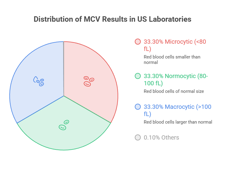
US laboratories sort results into three bands. Below 80 fL is microcytic. Between 80 and 100 fL is normocytic. Above 100 fL is macrocytic.
The Adult Reference Range and Why 80 to 100 Is Not Universal
Here is what surprises people. Not every American lab uses 80 to 100 fL. Some report an adult range of 79 to 97 fL, others 78 to 98 fL.
That means an MCV of 99 fL passes cleanly at one lab and gets flagged at another. Neither is wrong. Each sets its interval from its own analyzers, its own validation work, and its own local population.
How the Range Shifts With Age
Age moves the target substantially, and a pediatric result should never be judged against an adult chart.
MCV Reference Bands by Age Group
| Age group | Typical MCV range (fL) | What drives the difference |
| Newborn | 95 to 121 | Fetal hemoglobin and naturally large newborn cells |
| 6 months to 2 years | 70 to 86 | Rapid growth outpaces stored iron |
| 2 to 12 years | 73 to 87 | Gradual climb toward adult values |
| 12 to 18 years (boys) | 78 to 98 | Puberty and expanding blood volume |
| 12 to 18 years (girls) | 78 to 102 | Menstrual iron losses begin |
| Adults over 18 | 78 to 100 (lab dependent) | Stable unless disease or deficiency intervenes |
Parents booking pediatric panels with us often ask why a toddler’s MCV of 74 fL draws no alarm. At that age it sits comfortably inside the expected band.
Read Your Own Report, Not a Chart From the Internet
Every US laboratory prints its reference interval beside your result. That printed range is the only one that applies to your sample.
Judging your number against a range found online, rather than the one on your own report, produces far more anxiety than insight.
Table 1: The Three MCV Zones Side by Side
| Zone | MCV Range (fL) | Most Common USA Causes | Companion Markers to Check | Confirmatory Test to Request |
| Marked microcytosis | Below 70 | Thalassemia trait, long-standing iron deficiency | RBC count often high in thalassemia; RDW normal | Hemoglobin electrophoresis plus ferritin |
| Microcytosis | 70 to 79 | Iron deficiency, chronic inflammation, lead exposure | Ferritin low, RDW elevated, MCH low | Ferritin and transferrin saturation |
| Normocytic | 80 to 100 | Acute blood loss, kidney disease, anemia of inflammation | Hemoglobin, reticulocyte count, creatinine | Reticulocyte count and kidney panel |
| Borderline macrocytosis | 101 to 109 | Alcohol, liver disease, hypothyroidism, medications | Liver enzymes, TSH, B12, folate | B12, folate, TSH, liver panel |
| Marked macrocytosis | 110 and above | B12 or folate deficiency, myelodysplastic syndrome | Hypersegmented neutrophils, platelet count | B12, methylmalonic acid, peripheral smear |
| Normal MCV, high RDW | 80 to 100 with RDW above 14.5% | Combined iron plus B12 deficiency, early deficiency | Ferritin and B12 both required | Ferritin, B12, folate, peripheral smear |
Abnormal red cell values are common enough that most Americans encounter one eventually. CDC survey data covering August 2021 through August 2023 put anemia prevalence at 9.3% among people age 2 and older, with 13.0% in females against 5.5% in males, climbing to 12.5% in adults 60 and older.
Low MCV (Microcytosis): The Small-Cell Pattern
Small cells signal that the marrow ran short of hemoglobin-building material. Your body defends hemoglobin concentration inside each cell, so when supply falls it shrinks the cell rather than diluting it.
Iron Deficiency, the Most Common Cause in the USA
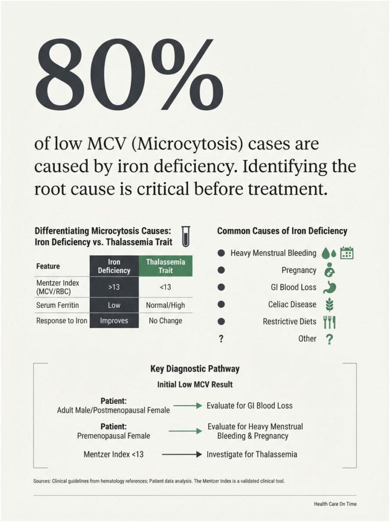
Iron deficiency dominates this category. Without adequate iron, hemoglobin production slows and the marrow releases smaller cells to hold concentration steady.
Frequent drivers include heavy menstrual bleeding, pregnancy, gastrointestinal blood loss, celiac disease, bariatric surgery, and restrictive eating patterns.
Why Ferritin Drops Before MCV Does
Ferritin is your iron savings account. It empties first. Serum iron falls next. Only afterward does red blood cell size begin to slide.
That sequence explains why someone can feel wrecked with a perfectly normal MCV blood test result. Cell shrinkage is a late signal, not an early alarm.
Thalassemia Trait and the Mentzer Index
Thalassemia trait also produces microcytosis, and it appears across Americans with Mediterranean, South Asian, Southeast Asian, Middle Eastern, and African ancestry. Mistaking it for iron deficiency wastes months and risks iron overload.
Running the Mentzer Calculation on Your Own Report
The Mentzer Index divides MCV in fL by RBC count in millions per microliter. Above 13 favors iron deficiency, where the marrow makes fewer and smaller cells. Below 13 favors thalassemia trait, where cell numbers stay normal but each cell is small.
Reported performance sits near 91% sensitivity and 83% specificity for iron deficiency in adults, though a pediatric study found much weaker figures at 74% sensitivity and 63% specificity for beta-thalassemia trait. Treat it as a pointer, never a verdict.
Chronic Inflammation, Lead, and the Less Common Causes
Long-running inflammation from rheumatoid arthritis, inflammatory bowel disease, or persistent infection can produce mild microcytosis by locking iron away from the marrow. Lead exposure and sideroblastic anemia complete the list.
Our lab partners report that inflammatory microcytosis usually stays milder than true iron deficiency, settling in the high 70s rather than the low 70s.
When Low MCV in an Adult Means Look for Bleeding
This subsection matters more than any other in the low-MCV discussion. In American men and postmenopausal women, new iron deficiency is treated as blood loss until proven otherwise, because those groups have no routine physiological reason to lose iron.
Physicians typically ask about black or bloody stools, unexplained weight loss, and changes in bowel habit. Depending on age and symptoms, colonoscopy or upper endoscopy follows.
Correcting the iron without finding the leak treats the number while the cause continues. That distinction has saved lives, and it is the reason clinicians push back when patients want a supplement instead of a workup.
High MCV (Macrocytosis): The Large-Cell Pattern
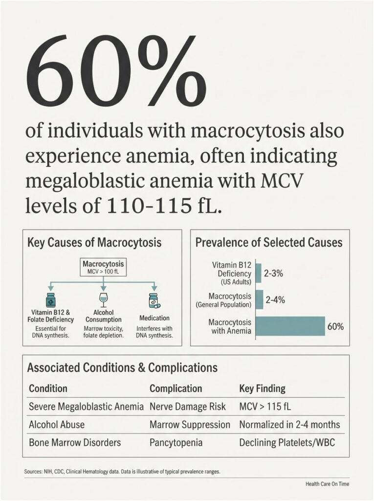
Large cells usually mean the marrow struggled to divide cells properly, so each one kept growing before splitting. Roughly 2% to 4% of people have macrocytosis, and about 60% of those also have anemia. An extremely high MCV in the 110 to 115 fL range often signals megaloblastic anemia.
B12 and Folate, the Deficiencies That Should Not Wait
Vitamin B12 and folate both drive DNA synthesis. Without them, red cell precursors expand while cell division stalls.
Vitamin B12 deficiency affects roughly 2% to 3% of US adults, with risk elevated by malabsorption, limited dietary intake, medications including metformin and proton pump inhibitors, and older age. A serum level below 180 pg/mL is diagnostic.
Why B12 Must Be Checked Before Folate Is Treated
Folic acid repairs the blood picture of B12 deficiency while nerve damage quietly continues underneath. Numbness, balance problems, and memory changes can become permanent.
This is the single most consequential sequencing error in the entire topic. Test both together, treat B12 first.
Alcohol, Liver Disease, and Thyroid
Alcohol raises MCV through direct marrow toxicity and by depleting folate. Elevated MCV appears in roughly 40% to 60% of chronic heavy drinkers, and because red cells live about 120 days, it takes two to four months to normalize after drinking stops.
Liver disease alters the lipid composition of red cell membranes, expanding surface area. Hypothyroidism slows marrow output and commonly nudges red blood cell size upward, often correcting once thyroid hormone is replaced.
Medications That Push MCV Up
Drugs that interfere directly with DNA synthesis include hydroxyurea, methotrexate, azathioprine, zidovudine, phenytoin, valproate, and multiple chemotherapy agents. These raise MCV within weeks of starting.
Drugs that work indirectly reduce nutrient absorption. Metformin and long-term proton pump inhibitors both lower B12 over months to years, producing macrocytosis that looks nutritional because it is.
In cases reviewed by our medical team, a complete medication and supplement list explains more borderline macrocytosis than any other single factor. Bring the actual bottles, not a remembered list.
When High MCV Points Toward the Bone Marrow
Myelodysplastic syndromes disrupt normal cell development and appear more often in older adults. A rising reticulocyte count also lifts MCV directly, since these immature cells run noticeably larger than mature red cells, which happens during recovery from bleeding or hemolysis.
The finding that raises clinical concern is not a high MCV by itself. It is macrocytosis alongside falling platelets or white cells.
The Normal MCV Trap: When an Average Hides Two Problems
MCV is an average. Averages conceal distributions, and this is where a normal-looking report misleads people for months.
What RDW Catches That MCV Misses
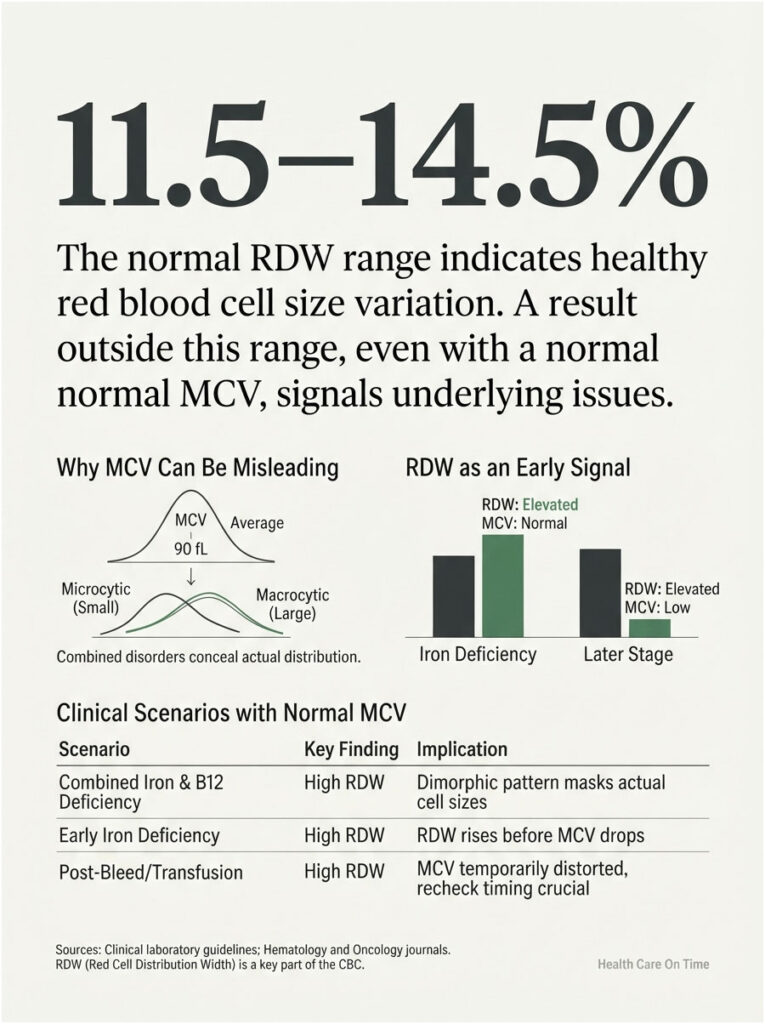
The Merck Manual states it plainly: a high RDW may be the only indication of simultaneous microcytic and macrocytic disorders, a combination that can produce a normal MCV because MCV reports only the mean.
RDW measures spread. Normal sits near 11.5% to 14.5%. Anything above that means your red cells are not uniform in size.
Combined Iron Plus B12 Deficiency
Picture half your cells shrunken from low iron and half enlarged from low B12. The average lands near 90 fL. Your MCV blood test reads normal.
Pathology references describe this dimorphic pattern directly, noting that MCV, MCH, and MCHC all reflect averages that fail to capture mixed red cell populations. A peripheral blood smear reveals what the indices hide.
Early Iron Deficiency, Before MCV Falls
RDW frequently rises before MCV drops, which makes it an earlier signal than the number most people watch.
Readers who contact HealthCareOnTime after months of unexplained fatigue with normal labs are often carrying an elevated RDW that nobody walked them through.
After Transfusion or During Recovery
A recent transfusion mixes donor cells with your own. Recovery from bleeding floods circulation with large reticulocytes.
Both scenarios distort MCV temporarily. Timing your recheck matters as much as ordering it.
Why the Same Person Gets Different MCV Results
This is the question standard explainers skip, and it has two distinct answers. Either your body changed, or the measurement changed.
How Stable Your MCV Should Actually Be
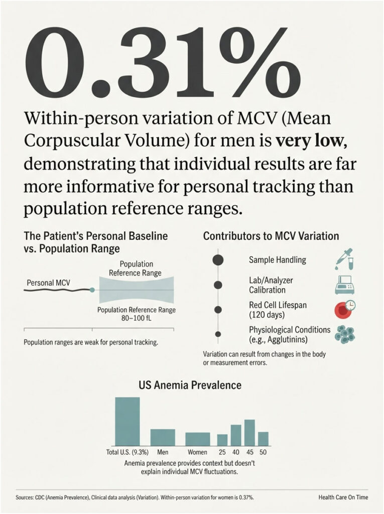
An analysis built on national US survey data collected between 1999 and 2002 found mean corpuscular volume had the lowest within-person coefficient of variation of any hematology test examined, at 0.31% for men and 0.37% for women, with an index of individuality of just 0.06.
Those figures come from an older survey cycle, so treat them as an order-of-magnitude guide rather than a precise current constant. The direction of the finding has held up in later work.
Your own MCV varies by well under one percent when nothing is wrong.
The Individuality Problem With Population Reference Ranges
An index of individuality that low means the population range is a weak tool for tracking you personally. Your own previous results carry far more information.
A move from 84 fL to 92 fL keeps you inside normal the entire journey. It is also close to a ten percent change in a value that should barely move.
The 120-Day Lag
Red blood cells circulate for roughly four months. MCV reflects every cell in circulation, including ones built long before your treatment started.
Start iron in January and your February MCV will look nearly unchanged. That is expected physiology, not treatment failure.
Sample Handling
Red cells absorb water during storage, which falsely raises MCV and lowers MCHC. Because hematocrit is derived from MCV, hematocrit can read high as well.
A tube that sat overnight at room temperature before reaching the analyzer reports differently than one run within the hour. The reverse also happens: drawing a small volume into a standard EDTA tube shrinks cells, since EDTA is hypertonic, producing a falsely low MCV and falsely high MCHC.
Different Lab, Different Analyzer, Different Reference Range
Impedance analyzers and optical analyzers size cells using different physics. Each manufacturer calibrates independently.
Layer a different reference interval on each report and you get two accurate results that disagree. Comparing them starts with comparing their ranges.
Conditions That Fool the Analyzer
Cold agglutinins make red cells clump, so the instrument counts a clump as one large cell and reports a falsely high MCV. Severe hyperglycemia causes osmotic swelling once cells enter the analyzer diluent.
Across panels processed through our diagnostic network, a wildly unexpected MCV is more often a handling or interference artifact than new disease.
Table 2: MCV and Anemia by the Numbers in the USA
| Metric | Figure | Population | Source |
| Anemia prevalence | 9.3% | US, age 2 and older | CDC/NCHS NHANES, Aug 2021 to Aug 2023 |
| Anemia by sex | 13.0% female vs 5.5% male | US, age 2 and older | CDC/NCHS NHANES Data Brief 519 |
| Anemia in older adults | 12.5% | US adults 60 and older | CDC/NCHS NHANES Data Brief 519 |
| Anemia in adolescent girls | 17.4% | US girls 12 to 19 | CDC/NCHS NHANES Data Brief 519 |
| Macrocytosis prevalence | 2% to 4%, about 60% also anemic | General population | Cleveland Clinic |
| Vitamin B12 deficiency | 2% to 3% | US adults | American Family Physician, Sept 2025 |
| MCV within-person variation | 0.31% men, 0.37% women | US NHANES 1999 to 2002 | NHANES biological variation analysis |
| CBC with differential order share | 7.26% of all US lab procedures | US CLIA labs, 2024 | Definitive Healthcare claims analysis |
What Happens Next After an Abnormal MCV
An out-of-range result starts a short, predictable investigation. Knowing the sequence lets you ask sharper questions.
The Follow-Up Tests Doctors Order
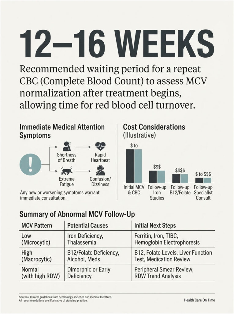
For low MCV: ferritin, serum iron, transferrin saturation, total iron-binding capacity, and hemoglobin electrophoresis when iron studies return normal.
For high MCV: serum B12, folate, TSH, liver panel, reticulocyte count, and a peripheral blood smear. Methylmalonic acid gets added when B12 sits in the borderline zone between roughly 180 and 300 pg/mL.
How Long to Wait Before a Repeat CBC Is Meaningful
Eight to twelve weeks is the standard interval after starting iron or B12. Testing sooner measures cells that were already finished.
Our medical reviewers suggest booking the recheck at the laboratory that ran the original panel. That removes analyzer differences from the comparison entirely.
Red-Flag Symptoms That Should Not Wait
Chest pain, severe shortness of breath, fainting, racing heartbeat, confusion, new numbness, tingling, or balance trouble all warrant prompt medical attention rather than a scheduled follow-up.
Blood in stool, black stools, or unusually heavy menstrual bleeding alongside a low MCV also move the timeline forward.
What Testing Costs and Where to Get It
MCV arrives inside a CBC, never as a standalone order. CPT code 85025 covers the CBC with automated differential, and it was the most commonly billed chemistry and hematology test in the United States during 2024, accounting for 7.26% of all lab procedures.
Because it is bundled, the marginal cost of getting your MCV blood test is effectively zero once a CBC is ordered. Most US insurance plans cover a CBC as part of preventive care, and direct-to-consumer panels are widely available without a physician order.
The follow-up tests carry separate costs. Ferritin, B12, folate, and TSH are inexpensive and routinely covered when an abnormal CBC justifies them, so ask your provider to order them together rather than sequentially.
Table 3: Your Result Pattern, Your Next Move
| If Your Pattern Looks Like This | Most Likely Explanation | What to Ask Your Doctor For | Reasonable Timeline |
| MCV under 80, RDW high, ferritin low | Iron deficiency anemia | Iron studies plus a discussion of blood loss source | Recheck CBC 8 to 12 weeks after treatment starts |
| MCV under 75, RDW normal, RBC count high | Thalassemia trait | Mentzer Index review and hemoglobin electrophoresis | One-time workup, no repeat CBC needed |
| MCV above 100 with numbness or balance change | B12 deficiency | Serum B12 and methylmalonic acid, urgent review | Same week |
| MCV 101 to 108, rest of CBC normal, no symptoms | Alcohol, medication, or benign variation | B12, folate, TSH, liver panel, medication review | Recheck in 3 months |
| MCV normal, RDW above 14.5%, ongoing fatigue | Mixed or early deficiency | Ferritin and B12 together, plus peripheral smear | Within 2 to 4 weeks |
| MCV shifted 5 fL or more from your own baseline | Real biological change | Repeat CBC at the same lab, then investigate | Repeat in 4 to 6 weeks |
| MCV differs between two labs the same week | Analyzer or sample handling difference | Compare each report’s printed reference range | No action beyond comparison |
| MCV above 110 with low platelets or white cells | Marrow-level problem | Hematology referral and peripheral smear | Within days |
Mistakes That Cost People Months
Four errors surface again and again, and each one delays a correct answer.
Starting Supplements Before the Cause Is Identified
Taking iron for a low MCV caused by thalassemia trait accomplishes nothing and risks iron overload. Taking it for iron deficiency caused by colon bleeding treats the number while the source keeps draining.
Find the cause first. The supplement is step two.
Treating Folate While a B12 Deficiency Hides Underneath
Folic acid normalizes the blood picture and masks the deficiency causing neurological damage. That is precisely why clinicians order both together.
Comparing Two Labs Without Comparing Their Reference Ranges
An MCV of 98 fL reads normal against 80 to 100 and abnormal against 79 to 97. Same cells, opposite verdict.
Before concluding your red blood cell size increased, confirm the reference interval did not simply narrow.
Chasing a One-Point Change
A move from 91 to 92 fL falls within ordinary analyzer imprecision.
Trends across three or four results tell a story. Single-point differences almost never do.
How to Track Your MCV Properly Over Time
Because MCV holds so steady within a person, it becomes a genuinely useful personal marker once you establish a baseline.
Build Your Own Baseline First
Pull your last three CBC reports and write down MCV, RDW, hemoglobin, and the reference range each lab used. That set is worth more than any published chart.
Your baseline is the number your MCV returns to when nothing is wrong.
Same Lab, Same Conditions, Same Question
Use one laboratory whenever possible. Draw at a similar time of day, in a similar hydration state, and confirm the sample reaches the analyzer promptly.
Consistency strips out the noise so the actual signal stands clear.
What to Bring to the Appointment
Bring prior CBC results, a full medication and supplement list, an honest account of alcohol intake, and any dietary restrictions.
Patients who arrive with that packet at HealthCareOnTime partner facilities typically shorten their workup by at least one visit.
Frequently Asked Questions
What does it mean if my MCV is high?
A high MCV means your red blood cells are larger than average, called macrocytosis. The usual explanations are vitamin B12 or folate deficiency, regular alcohol use, hypothyroidism, liver disease, and certain medications. It signals that further testing is warranted, not that a diagnosis has been made.
What does a low MCV blood test result mean?
A low MCV means smaller-than-average red blood cells, called microcytosis. Iron deficiency leads the list in the United States, followed by thalassemia trait and chronic inflammation. Doctors typically order ferritin and iron studies first, then consider hemoglobin electrophoresis if iron levels come back normal.
Is an MCV of 102 dangerous?
An MCV of 102 fL is mildly elevated and rarely dangerous alone, particularly when hemoglobin and the rest of your CBC look normal. Common explanations include alcohol, medications, and mild B12 or folate shortfalls. Testing B12, folate, TSH, and liver enzymes usually identifies the reason.
Can I have anemia with a normal MCV?
Yes. Normocytic anemia occurs with acute blood loss, kidney disease, and chronic inflammation. A normal MCV can also conceal combined iron plus B12 deficiency, where small and large cells average out. An elevated RDW alongside a normal MCV is the pattern that exposes it.
Why did my MCV change between two blood tests?
Either your marrow changed how it builds cells, or the measurement conditions changed. Storage time, a different analyzer, a different reference range, or a short draw volume can all shift the reported value. Real biological change typically takes weeks to months to become visible.
Does a high MCV mean cancer?
Rarely. Nutritional deficiencies, alcohol, thyroid problems, liver disease, and medications explain the overwhelming majority of high results. Bone marrow disorders such as myelodysplastic syndrome can raise MCV, but they generally disturb platelets and white cells too, not MCV in isolation.
Does alcohol raise MCV, and how fast does it come back down?
Yes. Alcohol raises MCV through direct marrow toxicity and folate depletion, and macrocytosis appears in roughly 40% to 60% of chronic heavy drinkers. Because red blood cells live about 120 days, MCV takes two to four months to normalize after drinking stops.
Can dehydration or stress change my MCV?
Dehydration concentrates blood and can lift hemoglobin and hematocrit, but it has little direct effect on average cell size. Short-term stress does not meaningfully move MCV. Values that swing sharply within days usually point to sample handling rather than physiology.
How long does it take for MCV to return to normal after starting iron?
Expect eight to twelve weeks before MCV shows clear improvement, and up to four months for full normalization. Reticulocytes rise within a week and hemoglobin responds within a month, which is why clinicians track those markers before rechecking red blood cell size.
Why is my Labcorp range different from my Quest range?
Each laboratory establishes reference intervals from its own analyzers, validation studies, and patient population. One may print 80 to 100 fL while another prints 79 to 97 fL. Always compare your result against the range on the report that produced it.
Can metformin cause a high MCV?
Indirectly, yes. Long-term metformin reduces vitamin B12 absorption, and the resulting deficiency raises MCV. Proton pump inhibitors act similarly. Anyone taking either medication long term should have B12 checked periodically rather than assumed adequate.
What foods actually change MCV?
Iron-rich foods such as red meat, lentils, spinach, and fortified cereals support normal red blood cell size when iron deficiency is the cause. B12 comes from meat, fish, eggs, dairy, and fortified products. Folate comes from leafy greens, beans, and fortified grains. Diet alone rarely corrects an established deficiency.
Medical Disclaimer
This article provides general education and does not replace professional medical advice, diagnosis, or treatment. MCV blood test results require interpretation by a licensed clinician alongside your full medical history, symptoms, medications, and the remainder of your complete blood count. Do not start or stop any supplement or medication based on this content. If you have concerning symptoms, contact a qualified healthcare provider promptly.
References
- Anemia Prevalence: United States, August 2021 to August 2023, CDC/NCHS Data Brief 519
- MCV Blood Test, Cleveland Clinic
- Macrocytosis: Causes, Symptoms and Treatment, Cleveland Clinic
- Evaluation of Anemia, Merck Manual Professional Edition
- Vitamin B12 Deficiency: Common Questions and Answers, American Family Physician
- Normal and Abnormal Complete Blood Count With Differential, StatPearls, NCBI Bookshelf
- Macrocytosis, StatPearls, NCBI Bookshelf
- Red Cell Indices, Clinical Methods, NCBI Bookshelf
- Biological variation of hematology tests based on the 1999 to 2002 NHANES, PubMed
- Mentzer Index Calculator, Medscape Reference
- Red Cell Distribution Width (RDW) Test, Medscape
- Blood Tests, National Heart, Lung, and Blood Institute
- Most common lab tests by procedure volume, Definitive Healthcare
- Unnecessary Testing, Harvard Medical School