
Your brain is the best-defended organ you own. Bone, three layers of membrane, and the blood-brain barrier turn away almost every germ that gets near it. So when bacteria manage to build a pocket of pus inside brain tissue, it usually means something else went wrong first, somewhere else in the body, and nobody caught it in time.
Table of Contents
Quick Answer: A brain abscess is a pocket of pus that forms inside brain tissue after bacteria, fungi, or parasites reach the brain and trigger inflammation. It’s a medical emergency because swelling raises pressure inside the skull. Brain abscess symptoms include a worsening headache, confusion, fever, one-sided weakness, and seizures. About 1,500 to 2,500 cases are diagnosed in the United States each year.
At a Glance
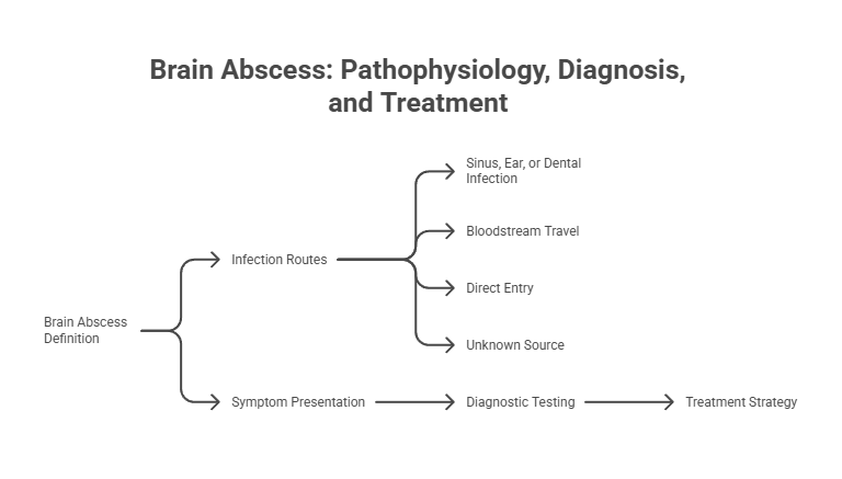
- A brain abscess, also called a cerebral abscess, is a walled-off collection of pus inside brain tissue rather than on its surface.
- Four routes get infection in: spread from a nearby sinus, ear, or dental infection; travel through the bloodstream; direct entry after head injury or surgery; and in roughly 15 percent of cases, a source nobody ever finds.
- Headache leads the symptom list at 69 to 70 percent, followed by mental status changes at 65 percent and focal neurological deficits at 50 to 65 percent.
- The textbook triad of fever, headache, and neurological deficit shows up together in fewer than half of patients, which is exactly why diagnosis gets delayed.
- MRI with contrast, especially diffusion-weighted imaging, is the test of choice and separates a cerebral abscess from a tumor with high reliability.
- Brain abscess treatment pairs 4 to 8 weeks of intravenous antibiotics with surgical drainage for most abscesses over 2 centimeters.
What a Brain Abscess Actually Is
Ask what is a brain abscess and the short answer is a localized area of dead and dying brain tissue, filled with pus, usually sealed off by a membrane. Clinicians use the broader term intracranial abscess when they want to include related infections that sit just outside brain tissue.
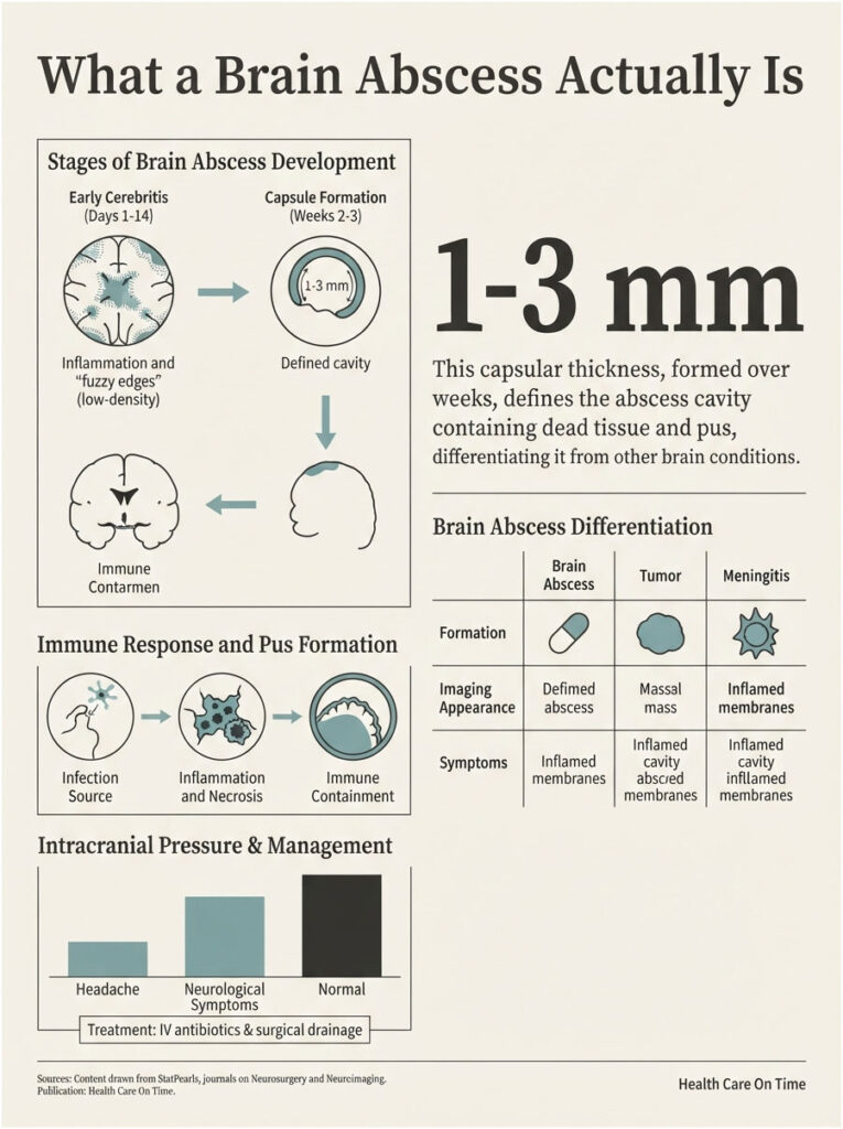
Here’s the part that surprises people. Your immune system builds the abscess on purpose.
White blood cells rush the invading bacteria, and the wreckage they leave behind, meaning dead cells, bacteria, and fluid, pools in one spot. Tissue then grows a wall around that pool to stop the infection from spreading. In a leg or a tooth, that containment strategy works beautifully. Inside a rigid skull, it creates an entirely new problem.
How Pus Forms Inside Brain Tissue
The process doesn’t start as an abscess. It starts as inflammation. Blood vessels widen, fluid leaks into nearby tissue, and a poorly defined zone of infected brain develops without any clear border.
Only later does the center liquefy into true pus. Our medical reviewers note that this two-stage progression explains something patients find confusing: an early scan can look ambiguous, and a repeat scan seven days later can tell a completely different story.
The Stages, From Cerebritis to Encapsulation
Early Cerebritis (Days 1 to 14)
For the first week or two, the lesion has fuzzy edges. Vascular congestion and swelling dominate, and imaging may show nothing more than a vague low-density patch.
This is the window where antibiotics alone have their best shot, because no thick wall is blocking the drug yet.
Capsule Formation (Weeks 2 to 3)
Around the two to three week mark, necrosis and liquefaction produce a defined cavity. A capsule forms with three layers: granulation tissue inside, collagen in the middle, an astroglial layer outside.
That wall usually measures 1 to 3 millimeters thick. Harvard Health reports a brain abscess can become fully formed in roughly two weeks, which is fast for something this dangerous.
Why the Skull Turns a Small Infection Into a Big Problem
Your skull can’t expand. Every millimeter the abscess claims has to come from somewhere, and the surrounding brain swells on top of that.
The result is increased intracranial pressure, which drives headache, vomiting, drowsiness, and eventually loss of consciousness. Pressure also squeezes blood vessels, starving tissue that was perfectly healthy an hour earlier.
Brain Abscess Compared With Look-Alike Conditions
Readers write to us regularly asking how a brain abscess differs from meningitis or a tumor, since all three can produce headaches and neurological changes. The distinction matters enormously, because the treatments have almost nothing in common.
| Feature | Brain Abscess | Subdural Empyema | Bacterial Meningitis | Brain Tumor |
| Anatomical location | Inside brain tissue (parenchyma) | Between the dura and arachnoid membranes | In the fluid and membranes surrounding the brain | Inside or adjacent to brain tissue |
| What collects there | Walled-off pus with a 1 to 3 mm capsule | Pus spreading across a membrane surface | Inflammatory cells in cerebrospinal fluid | Abnormal cell growth, no pus |
| Typical onset speed | Days to 2 weeks in about two-thirds of cases | Rapid, often within days | Hours to 2 days | Weeks to months |
| Hallmark imaging finding | Ring-enhancing lesion with restricted diffusion on DWI | Crescent-shaped collection along the dura | Often normal on CT; diagnosis rests on spinal fluid | Ring enhancement without restricted diffusion |
| Classic symptom pattern | Localized worsening headache, focal deficit, sometimes fever | Fever, headache, rapid neurological decline, seizures | Fever, severe headache, stiff neck, light sensitivity | Progressive deficit, morning headache, personality change |
| First-line management | IV antibiotics plus surgical drainage in most cases | Urgent surgical drainage plus IV antibiotics | IV antibiotics, no drainage needed | Biopsy, then surgery, radiation, or chemotherapy |
How Common Brain Abscesses Are in the United States
Brain abscesses are uncommon, and that rarity is part of why they slip past clinicians. A busy emergency physician might see a handful across an entire career.
National Case Estimates
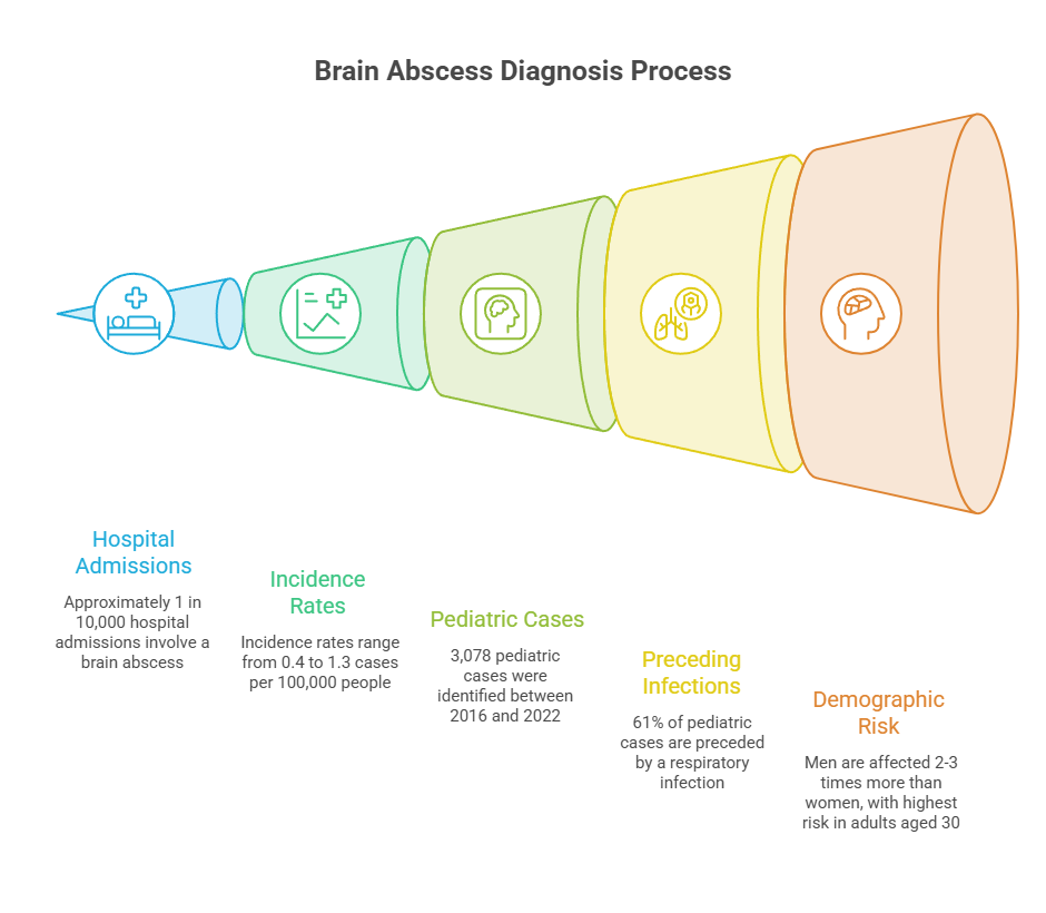
Roughly 1,500 to 2,500 cases are diagnosed annually in the United States, according to StatPearls via the National Library of Medicine. That’s about 1 in every 10,000 hospital admissions.
Published incidence estimates cluster between 0.4 and 1.3 cases per 100,000 people per year. Rates have been climbing over recent decades, particularly among older adults.
What CDC Surveillance Data Reveals
The richest American dataset came from a CDC investigation launched in 2022. Between January 2016 and May 2022, CDC identified 3,078 pediatric cases of brain abscess, epidural empyema, or subdural empyema across 40 children’s hospitals.
Monthly counts ran from 20 to 68, with a median of 38. Cases dropped below that median for 15 straight months after March 2020, then climbed through summer 2021 to a March 2022 peak before returning to baseline.
CDC also issued a national call for cases and collected 81 confirmed pediatric reports. Among those children, 61 percent had a respiratory infection in the six weeks before hospitalization. Sinusitis was the single most common at 26 percent.
Who Gets Them Most Often
Men are affected roughly two to three times as often as women. Adults in their thirties carry the highest adult risk, while pediatric cases cluster between ages 4 and 7. Newborns form a third at-risk group.
| Metric | Figure | Population or Setting | Source |
| Annual US cases diagnosed | 1,500 to 2,500 | All ages, nationwide | StatPearls / NLM, 2024 |
| Share of US hospitalizations | About 1 in 10,000 | Inpatient admissions | Cleveland Clinic Journal of Medicine |
| Pediatric cases logged | 3,078 | 40 US children’s hospitals, Jan 2016 to May 2022 | CDC MMWR 71(37), 2022 |
| Median pediatric patient age | 8 years (IQR 1 to 13) | Pediatric Health Information System | CDC MMWR 71(37), 2022 |
| Male share of pediatric cases | 65.1 percent | Pediatric Health Information System | CDC MMWR 71(37), 2022 |
| Preceding respiratory infection | 61.0 percent | 81 reported pediatric cases, 2021 to 2022 | CDC MMWR 71(37), 2022 |
| Median hospital stay | 10 days (IQR 6 to 21) | 71 discharged pediatric patients | CDC MMWR 71(37), 2022 |
One county-level example shows how sharply numbers can swing. Clark County, Nevada averaged four pediatric cases per year from 2015 through 2021. In 2022 it recorded 18, which triggered a health district advisory.
What Causes a Brain Abscess
Bacteria cause the large majority. Fungi and parasites account for a smaller share, mostly in people whose immune systems are already compromised.
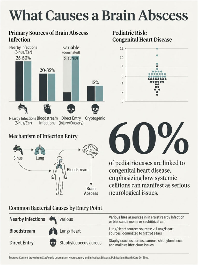
The route matters more than the organism does, because the entry point predicts where in the brain the abscess lands and which bacteria turn up in culture.
Spread From a Nearby Infection
Direct spread from a neighboring infection drives somewhere between 25 and 50 percent of cases. The infection erodes through thin bone or travels along small veins into brain tissue.
Sinus Infections and the Frontal Lobe
Paranasal sinus infections account for 30 to 50 percent of cases, which makes them the leading contiguous source by a wide margin. Frontal and ethmoid sinus infections tend to seed the frontal lobes, since barely any bone separates the two.
Patients booking imaging and lab work through HealthCareOnTime often ask when a sinus infection stops being routine. Our medical reviewers point to a simple marker: one that keeps worsening past two weeks, especially with forehead or eye swelling, deserves imaging rather than another antibiotic refill.
Ear and Mastoid Infections
Middle ear infections account for roughly 5 percent of cases. Because of the anatomy, ear and mastoid infections usually produce abscesses in the temporal lobe or the cerebellum.
Chronic, undertreated ear infections carry far more risk than one acute episode that clears on schedule.
Dental Infections and Oral Bacteria
Untreated dental abscesses and advanced gum disease send oral bacteria toward the frontal lobe. Case series report an average gap of about 18 days between a dental flare-up or procedure and the first neurological symptoms.
Here’s the trap. Fewer than half of patients with an odontogenic brain abscess still have any mouth pain by the time they reach a neurologist, so the connection gets missed.
Spread Through the Bloodstream
Hematogenous seeding explains another 20 to 35 percent. Bacteria enter the circulation from a distant infection and lodge in brain tissue, typically at the gray-white matter junction.
Heart and Lung Sources
The lungs are the most frequent distant source. Lung abscesses, empyema, bronchiectasis, and cystic fibrosis all raise risk.
Bacterial endocarditis is another documented route. So are pulmonary arteriovenous malformations, and roughly 10 percent of people with that condition develop a brain abscess at some point.
Bloodstream-driven cases tend to produce multiple abscesses rather than one.
Congenital Heart Disease in Children
In children, cyanotic congenital heart disease accounts for over 60 percent of cases. A right-to-left shunt lets blood skip the lungs, which normally filter circulating bacteria out.
Chronic low oxygen compounds it, leaving small brain regions poorly perfused and easier to colonize.
Direct Entry After Head Injury or Surgery
Penetrating head wounds, skull fractures, facial trauma, and neurosurgical procedures hand bacteria a direct path. Retained metal fragments or other foreign material can seed an infection months down the line.
Post-surgical cases skew hard toward staphylococci. An English cohort covering 2012 to 2023 found Staphylococcus aureus caused 27.8 percent of post-surgical brain abscesses.
When No Source Is Ever Identified
In at least 15 percent of cases, nobody ever finds the origin. Clinicians call these cryptogenic, and treatment moves ahead on empiric antibiotic coverage while cultures run.
The Bacteria Most Often Responsible
A meta-analysis pooling 9,699 patients from 123 studies identified streptococci and staphylococci as the dominant pathogens. Community-acquired cases lean toward oral cavity bacteria, including anaerobes.
Among the 81 pediatric cases CDC characterized, Streptococcus intermedius appeared in 46.1 percent, Streptococcus anginosus in 18.4 percent, Streptococcus pneumoniae in 11.8 percent, and Streptococcus constellatus in 9.2 percent. About 21 percent of specimens were polymicrobial.
Brain Abscess Symptoms and How They Develop
Brain abscess symptoms come from three overlapping problems: the infection itself, rising pressure inside the skull, and damage to whichever brain region the abscess has taken over.
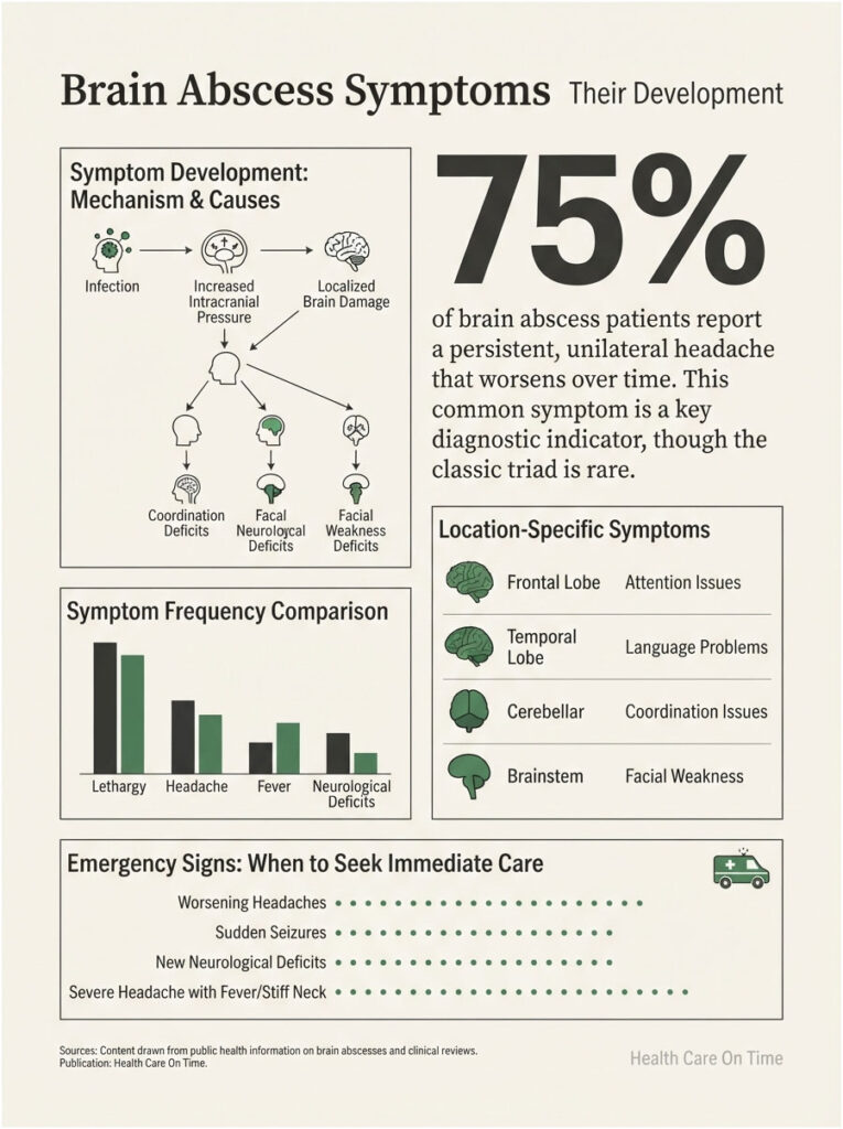
That mix produces a presentation vague enough to pass for migraine, flu, or a stubborn sinus headache. In roughly two-thirds of patients, symptoms have been running two weeks or less at diagnosis, and the average gap between first symptom and diagnosis is about eight days.
The Headache That Doesn’t Behave Normally
More than 75 percent of people with a brain abscess develop a dull, aching headache, and for many it’s the only symptom early on. Two features set it apart.
First, the pain tends to stay on the same side as the abscess instead of wrapping around the whole head. Second, it keeps escalating, and over-the-counter pain relievers barely touch it.
A headache that worsens steadily over days, resists medication, and stays parked in one spot is a different animal from one that comes and goes.
The Classic Triad and Why It Misleads
Medical training teaches a triad: fever, headache, focal neurological deficit. All three show up together in fewer than half of patients, and some series put the figure closer to 20 percent.
Fever is the most common no-show. Only 45 to 53 percent of patients run a temperature, meaning about half don’t. Waiting for a fever before taking a headache seriously costs days that matter.
Symptom Frequency, Ranked
Across published clinical series, brain abscess symptoms appear at these rates:
- Headache: 69 to 70 percent, usually localized to the affected side
- Mental status changes: 65 percent, ranging from mild confusion toward coma
- Focal neurological deficits: 50 to 65 percent, typically arriving days to weeks after the headache starts
- Fever: 45 to 53 percent, and often low-grade rather than high
- Nausea and vomiting: about 40 percent, driven by rising intracranial pressure
- Seizures: 25 to 35 percent, and sometimes the very first sign
- Stiff neck: about 15 percent, more common when the abscess leaks toward the ventricles
Lethargy sliding toward coma signals severe swelling. Clinicians treat it as a poor prognostic sign.
How Symptoms Shift Depending on Abscess Location
Where the abscess sits determines what breaks. A frontal lobe abscess brings headache, inattention, drowsiness, speech trouble, and generalized seizures.
Temporal lobe abscesses tend toward language problems and visual field defects. A cerebellar abscess produces unsteadiness, abnormal eye movements, vomiting, and lost coordination.
Brainstem abscesses, which are rarer, cause facial weakness, swallowing difficulty, and one-sided weakness. Frontal and temporal regions are the most frequent sites overall.
Symptoms in Children and Infants
Children present less predictably than adults do. CDC’s pediatric series found sinusitis in 61 percent of hospitalized children, skull osteomyelitis in 31.2 percent, and bacterial meningitis in 26 percent.
In infants, a bulging fontanelle, poor feeding, irritability, and expanding head circumference may be the only clues. A seizure in a previously healthy child always warrants urgent evaluation.
Changes That Call for Emergency Care
The pattern that repeats most often in published case reports is a person who felt unwell for several days, assumed it would pass, and only sought care once something neurological appeared.
| Scenario | What It May Point Toward | Recommended Action |
| Headache worsening daily for a week, fixed to one side, unrelieved by OTC medication | Rising intracranial pressure from a space-occupying lesion | Contact a physician the same day and request imaging, not another pain reliever |
| Sinus infection that worsens after 10 to 14 days, plus forehead or eye swelling | Possible spread beyond the sinus cavity | Seek same-day medical evaluation; imaging is typically warranted |
| Untreated dental abscess plus new headache, confusion, or facial swelling | Possible spread of oral bacteria | Go to an emergency room rather than waiting for a dental appointment |
| Sudden seizure in someone with no seizure history | Cortical irritation from an intracranial process | Call 911; seizures can be the first sign of a brain abscess |
| New weakness, numbness, or speech difficulty on one side of the body | Focal neurological deficit | Call 911 immediately; this overlaps with stroke presentation |
| Fever plus stiff neck plus severe headache | Meningitis or an abscess leaking toward the ventricles | Call 911; both are medical emergencies |
| Abrupt severe worsening of an existing headache with vomiting and confusion | Possible abscess rupture into the ventricular system | Call 911; this is a life-threatening complication |
Who Faces the Highest Risk
Most people who get a sinus or dental infection never come anywhere near a brain abscess. But certain conditions shift the odds enough to change how aggressively an infection should be handled.
Weakened Immune Systems
HIV infection, organ transplantation, active cancer, chemotherapy, and long-term corticosteroid use all raise risk. Immunocompromised patients also face a wider organism list, including fungi like Aspergillus and parasites like Toxoplasma gondii.
Fungal cases have risen alongside broader use of broad-spectrum antibiotics and immunosuppressive drugs.
Structural Heart and Lung Conditions
Cyanotic congenital heart disease, bacterial endocarditis, and pulmonary arteriovenous malformations bypass or overwhelm the body’s normal bacterial filtering. Bronchiectasis and cystic fibrosis add risk through repeated lung infection.
Recent Neurosurgery or Penetrating Head Injury
Any procedure or injury that breaches the skull opens a route. Shunt hardware carries ongoing risk, and infected shunts sometimes have to come out and be replaced.
Untreated Dental and Sinus Disease
CDC’s pediatric data found 44.9 percent of reported cases occurred in children with an underlying health condition, asthma being the most common at 11.5 percent. One detail deserves attention: 81.8 percent of those families had already sought outpatient care before hospitalization, which shows how easily an early presentation gets filed under something routine.
How Doctors Diagnose a Brain Abscess
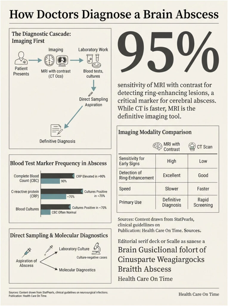
Diagnosis runs imaging first, laboratory work second, direct sampling third. Order matters here, because one common test can actually be dangerous.
Why Imaging Comes First
Symptoms alone can’t separate a cerebral abscess from a tumor, a stroke, or meningitis. Imaging settles it fast and reveals size, number, and location, all of which shape the treatment plan.
MRI With Contrast and Diffusion-Weighted Imaging
The 2024 ESCMID guideline, now widely referenced by US infectious disease specialists, recommends MRI for diagnosis with a strong recommendation backed by high-quality evidence. Contrast-enhanced MRI reveals the characteristic ring-enhancing lesion around a necrotic center.
Diffusion-weighted imaging is the detail that clinches it. Pus restricts water movement, so an abscess lights up bright on DWI while most tumors don’t.
MRI also catches early cerebritis, satellite lesions, and brainstem involvement that CT misses entirely.
When a CT Scan Is Used Instead
CT is faster and available in every American emergency department at 2 a.m., so that’s often where the workup starts. Contrast-enhanced CT shows a ring-enhancing lesion with surrounding swelling once the abscess has established itself.
Early cerebritis on CT may appear only as a vague low-density patch. A normal-looking early scan doesn’t close the case.
Blood Tests That Support the Picture
Standard workup includes a complete blood count with differential, erythrocyte sedimentation rate, C-reactive protein, and at least two sets of blood cultures drawn before antibiotics start where possible.
Our lab partners report that inflammatory markers in brain abscess behave inconsistently. One published series found 26 percent of patients had a normal CRP and 49 percent showed no elevated white cell count on admission. Normal bloodwork never rules this out.
Why Lumbar Puncture Is Usually Avoided
A spinal tap rarely helps here and can do real harm. Draining fluid from below a mass lesion can let swollen brain tissue shift downward, a potentially fatal complication.
If a lumbar puncture is considered at all, imaging comes first to rule out significant mass effect.
Aspiration and Laboratory Culture
Sampling the pus directly identifies the organism and steers antibiotic selection. Material goes for Gram stain, culture, histopathology, and molecular testing.
Current guidance recommends molecular diagnostics when cultures come back negative, which happens often once antibiotics are already running.
How Brain Abscesses Are Treated
Brain abscess treatment combines drugs and, for most patients, surgery. All of it happens in a hospital, with the early phase in intensive care for sicker patients.
Emergency Stabilization
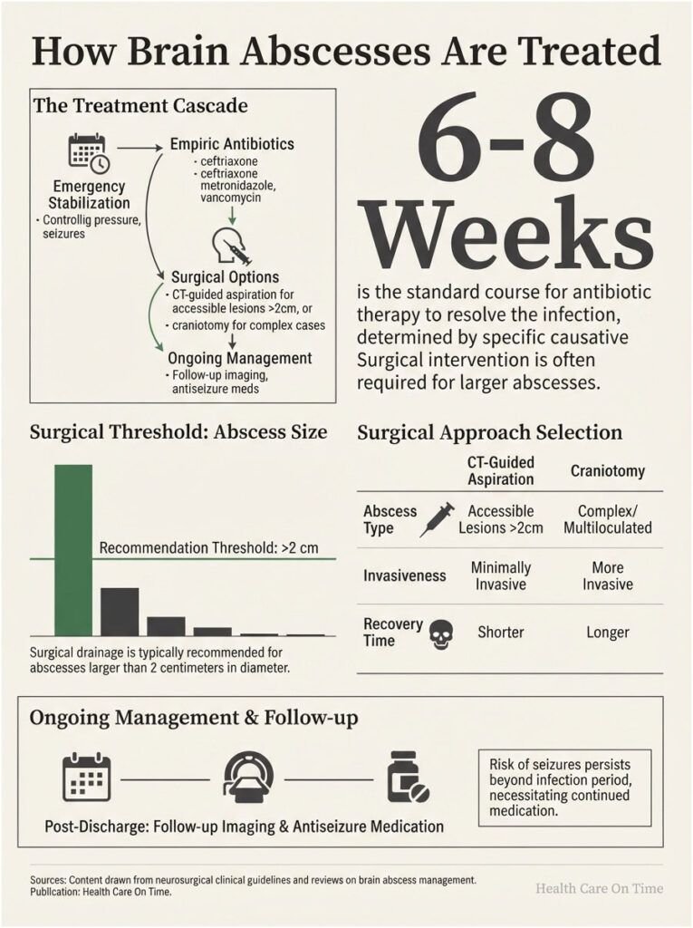
First priority is controlling intracranial pressure and stopping seizures. Dexamethasone may be given short-term to cut swelling, though it’s used selectively, since steroids can blunt the immune response and slow antibiotic penetration.
Antiseizure medication is standard, given that a quarter to a third of patients seize.
Antibiotic Therapy and How Long It Runs
Empiric coverage typically starts with a third-generation cephalosporin like ceftriaxone or cefotaxime, plus metronidazole for anaerobes and vancomycin when staphylococci are in play. CDC’s pediatric series recorded vancomycin in 91.2 percent of cases, ceftriaxone in 88.8 percent, and metronidazole in 81.2 percent.
Post-neurosurgical cases follow a different path. Current guidance recommends a carbapenem combined with vancomycin or linezolid.
Brain abscess treatment runs long. Guidelines recommend 6 to 8 weeks of antimicrobial therapy, and most American protocols work within a 4 to 8 week range depending on response.
Some antibiotics are poor choices no matter what grows. First-generation cephalosporins, aminoglycosides, and tetracyclines cross the blood-brain barrier badly and get skipped.
When Surgery Becomes Necessary
Guidelines recommend aspiration or excision whenever it’s feasible, with toxoplasmosis as the main exception. Drainage relieves pressure and delivers a specimen for culture at the same time.
The 2 Centimeter Threshold
Abscesses over 2 centimeters are generally considered for drainage. Smaller, deep-seated lesions may be managed with antibiotics alone under close imaging follow-up.
Other triggers for surgery include worsening pressure, failure to shrink on antibiotics, gas inside the cavity, and any abscess at risk of rupturing.
Aspiration Versus Craniotomy
CT-guided or stereotactic needle aspiration is the less invasive route and works well for accessible single lesions. Burr hole drainage plays a similar role.
Craniotomy, which means opening the skull, gets reserved for multiloculated abscesses, lesions that fail aspiration, or cases where complete removal is simply safer.
Managing Swelling and Seizure Risk
Follow-up imaging tracks whether the cavity is closing, often weekly during the treatment course. Antiseizure medication frequently continues after discharge, because seizure risk outlasts the infection.
Outlook, Recovery, and Long-Term Effects
Before antibiotics and modern imaging, a brain abscess was close to uniformly fatal. That’s no longer the picture.
Survival Figures Under Modern Care
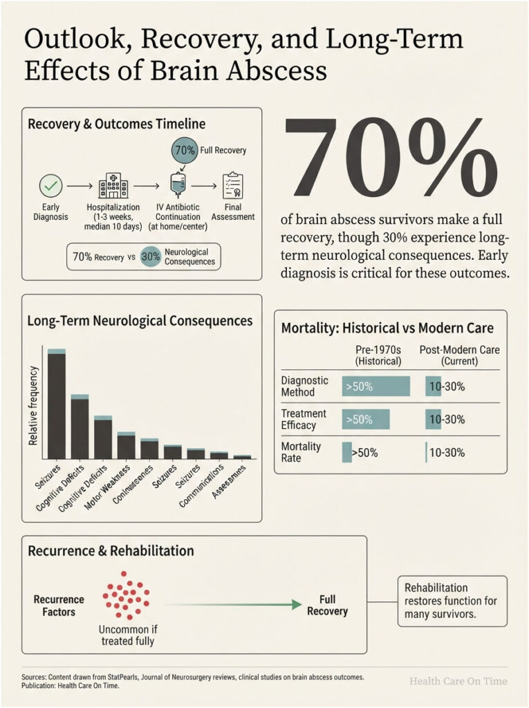
Modern cohorts report mortality around 15 to 20 percent, and MedlinePlus places the range at 10 to 30 percent across all cases. Larger population studies have found 30-day mortality near 15 percent and one-year mortality near 21 percent.
Children do better than adults. Certain groups do worse, with mortality reported as high as 50 percent after solid organ transplantation.
Early diagnosis remains the single biggest factor anyone can actually change. Rupture into the ventricular system is still the most dangerous complication.
What Recovery Looks Like, Week by Week
Hospitalization typically runs one to three weeks, with a median stay of 10 days in CDC’s pediatric data. IV antibiotics continue for weeks afterward, often through a PICC line at home or at an infusion center.
Repeat scans confirm the cavity is closing. Fatigue hits hard through the first month and frequently lingers for several more.
Lasting Neurological Effects
Around 70 percent of patients recover fully. The other 30 percent live with some neurological consequence, ranging from mild to significant.
Epilepsy develops in roughly 30 percent of survivors. Weakness, speech difficulty, cognitive changes, and mental fatigue also show up, and rehabilitation restores meaningful function for many people.
PET imaging research has shown that larger abscesses leave larger zones of reduced brain activity years later. That’s one more argument for moving fast.
How Often a Brain Abscess Returns
Recurrence is uncommon when the full antibiotic course gets completed and the original source is fixed. Leaving the underlying sinus, ear, or dental problem untreated is the most common reason one comes back.
Lowering Your Risk
You can’t prevent every case. Several habits meaningfully move the odds.
Treating Infections Before They Travel
Sinus infections, ear infections, and skin infections that aren’t improving on a reasonable course of treatment deserve reassessment, not patience. Delay is what gives bacteria the runway.
Dental Care as Brain Protection
Untreated cavities and gum disease supply the oral bacteria found in a large share of community-acquired cases. Our medical reviewers frame routine dental care as neurological protection, not cosmetic upkeep.
Antibiotic Prophylaxis for Certain Heart Conditions
People with specific congenital heart conditions or prosthetic valves may be told to take antibiotics before dental work. That call belongs to your cardiologist, who weighs your anatomy against current American Heart Association guidance.
Wound and Post-Surgical Care
Follow incision care instructions after any head or facial surgery. Drainage, redness, or fever after a neurosurgical procedure should be reported the same day, not at the next scheduled visit.
Frequently Asked Questions
Can a tooth infection cause a brain abscess?
Yes, though it’s rare. Oral bacteria from an untreated dental abscess can travel through nearby tissue or the bloodstream and seed the frontal lobe. Published series report an average of about 18 days between the dental problem and neurological symptoms, and fewer than half of those patients still have mouth pain by then.
How long does it take for a brain abscess to form?
Roughly two weeks to become fully formed. The first one to two weeks involve inflammation without a defined border, then a capsule develops around the pus over the following one to two weeks. Symptoms usually appear before that capsule finishes forming.
What is the survival rate for a brain abscess?
Modern cohorts report mortality between 10 and 30 percent, with most landing near 15 to 20 percent. Survival is better in children and worse in transplant recipients and other immunocompromised patients. Early diagnosis and prompt drainage are the strongest predictors of a good outcome.
Can a sinus infection lead to a brain abscess?
Paranasal sinus infections account for 30 to 50 percent of brain abscess cases, making them the leading contiguous source. Only thin bone separates the frontal and ethmoid sinuses from the frontal lobes. A sinus infection worsening past two weeks, especially with forehead or eye swelling, warrants prompt evaluation.
Is a brain abscess the same as meningitis?
No. A brain abscess is a walled-off pocket of pus inside brain tissue, while meningitis is inflammation of the membranes and fluid surrounding the brain. Meningitis is treated with antibiotics alone; most brain abscesses also need surgical drainage. Both are medical emergencies.
Can a brain abscess be mistaken for a brain tumor?
Yes, because both can appear as ring-enhancing lesions on contrast imaging. Diffusion-weighted MRI resolves it in most cases, since pus restricts water movement and appears bright while most tumors don’t. Direct sampling confirms the diagnosis when imaging stays unclear.
Do all brain abscesses require surgery?
No. Abscesses under 2 centimeters, those buried in critical brain regions, and cases with multiple small lesions may be managed with antibiotics alone under close imaging follow-up. Current guidelines still recommend aspiration or excision whenever it can be done safely.
What antibiotics are used to treat a brain abscess?
Empiric therapy usually pairs a third-generation cephalosporin such as ceftriaxone with metronidazole, adding vancomycin when staphylococci are suspected. Post-surgical cases typically get a carbapenem plus vancomycin or linezolid. Regimens narrow once cultures identify the organism. First-generation cephalosporins, aminoglycosides, and tetracyclines penetrate the brain poorly.
Can you fully recover from a brain abscess?
About 70 percent of patients recover fully. The remaining 30 percent experience some lasting neurological effect, with epilepsy occurring in roughly 30 percent of survivors. Recovery depends heavily on how early treatment began, plus abscess size and location. Rehabilitation restores meaningful function for many patients.
Are brain abscesses contagious?
No. A brain abscess can’t pass from person to person. The bacteria involved, including Streptococcus and Staphylococcus species, are common organisms that often already live in your mouth, skin, or airways. An abscess forms only when they reach brain tissue through a specific route.
Will seizures happen after a brain abscess?
Seizures occur in 25 to 35 percent of patients during the acute illness and can be the first sign. Longer term, roughly 30 percent of survivors develop epilepsy. Many patients continue antiseizure medication after discharge, with duration determined by their neurologist.
What does a brain abscess look like on an MRI?
It appears as a ring-enhancing lesion, meaning a bright circular rim around a darker center, surrounded by swelling. On diffusion-weighted imaging the center appears bright because pus restricts water movement. The rim usually measures 1 to 3 millimeters and looks smooth and even.
Medical Disclaimer: This article is for informational purposes only and does not constitute medical advice, diagnosis, or treatment. A brain abscess is a medical emergency. If you or someone near you develops a severe or worsening headache, confusion, a seizure, sudden weakness, or a stiff neck with fever, call 911 or go to the nearest emergency room. Always consult a qualified healthcare provider about your specific situation.
References
- Brain Abscess, StatPearls, National Library of Medicine
- Pediatric Brain Abscesses, Epidural Empyemas, and Subdural Empyemas Associated with Streptococcus Species, CDC MMWR 71(37)
- Increase in Pediatric Intracranial Infections During the COVID-19 Pandemic, CDC MMWR 71(31)
- Brain Abscess, Cleveland Clinic Health Library
- Cerebral Abscess, Johns Hopkins Medicine
- Brain Abscess, Harvard Health Publishing
- Abscess of the Brain, Merck Manual Consumer Version
- Brain abscess, MedlinePlus, National Library of Medicine
- ESCMID guidelines on diagnosis and treatment of brain abscess in children and adults, Clinical Microbiology and Infection